Abstract
Odontomas are nonaggressive, hamartomatous developmental malformations composed of mature tooth substances and may be compound or complex depending on the extent of morphodifferentiation or on their resemblance to normal teeth. Among them, complex odontomas are relatively rare tumors. They are usually asymptomatic in nature. Occasionally, these tumors become large, causing bone expansion followed by facial asymmetry. Odontoma eruptions are uncommon, and thus far, very few cases of erupted complex odontomas have been reported in the literature. Here, we report the case of an unusually large, painless, complex odontoma located in the right posterior mandible.
A complex odontoma is a hamartomatous lesion or malformation of odontogenic origin in which both epithelial and mesenchymal cells exhibit complete differentiation and all the dental tissues are represented. The individual hard tissues are well developed but in a more or less disorderly pattern.1
Although the etiology of complex odontomas is not clearly known, several theories have been proposed, which include trauma, infection, family history, and genetic mutation. Such odontomas may be discovered at any age, but the age with the greatest prevalence is the second decade of life.2 These tumors have a slight male predilection and are commonly seen in the posterior mandible.3,4 Complex odontomas are mostly asymptomatic in nature and are usually found in routine radiographic examinations.2
Complex odontoma rarely erupts into the oral cavity. The eruption of this lesion differs from tooth eruption because the lesion has no periodontal ligament; this could be attributed to bone sequestration or the remodeling of jaw bones.2 Eruption causes pain, inflammation, and infection of the adjacent structures. Thus far, a total of 11 cases of erupted complex odontomas have been reported in the literature.3 Garcia-Consuegra et al. reported pain and inflammation in association with a odontoma in only 4% of Spanish patients.5 Thus far, to the best of our knowledge, there have been no reports of an erupted complex odontoma that led to ulceration on the buccal mucosa. However, the odontoma in the present case measured 3.5 cm mesiodistally and 4 cm buccolingually and is rare. Vengal et al. reported a large erupted complex odontoma that measured 3 cm mesiodistally and 2 cm buccolingually.2 Dua et al. reported an erupted complex odontoma, which had a maximal dimension of 2.5 cm.3 The weight of the largest reported odontoma was 0.3 kg.6 However, the large complex odontoma in the present case weighed 43.5 g and was thus unusually large. This type of lesion is usually asymptomatic in nature when small, and when it increases in size, it expands the jaw and causes facial asymmetry, as in the present case. Therefore, here, we report this interesting case of a large erupted complex odontoma, which was associated with the expansion of the cortical plates, facial asymmetry, and the ulceration of the buccal mucosa.
A 22-year-old male presented to the Department of Oral Medicine and Radiology, ITS Dental College, Murad Nagar, Ghaziabad, Uttar Pradesh, India, with the chief complaint of swelling on the lower right side of his face over the past 6 years. The condition started with a mild, dull, and intermittent pain in this tooth region, which regressed after taking medication; this was followed by a small asymptomatic swelling that gradually increased in size over the course of 6 years. Initially, the patient underwent no treatment due to the asymptomatic nature of his medical condition.
Extraoral examination revealed gross facial asymmetry with swelling present on the right lower-third region of the patient's face (Fig. 1). The swelling extended anteroposteriorly from 2 cm posterior to the right corner of the mouth to the posterior border of the ramus, and superoinferiorly from 1 cm below the ala-tragus line to the lower border of the mandible. There were no secondary signs on the overlying skin.
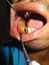
Intraoral examination revealed a yellowish brown, irregularly shaped solid mass, appearing as calculus, measuring approximately 3.5 cm (mesiodistally)×4 cm (buccolingually), situated in the region of the right posterior mandibular teeth. The mass extended mesiodistally from the region of the second premolar to that of the third molar. Buccally, the vestibular space was encroached upon by the mass, causing tissue tags on the buccal mucosa at the occlusal level due to repeated cheek biting (Fig. 2). The right mandibular second premolar and the first, second, and third molars were clinically missing. An ulcer was observed on the buccal mucosa adjacent to the lesion, which was due to chronic irritation caused by the lesion that started bleeding upon manipulation. The patient was able to occlude his teeth properly. Upon palpation, we found that this lesion was bony hard, mobile, and nontender in nature. Based on the abovementioned clinical findings, a provisional diagnosis of fibro-osseous lesion, probably cemento-ossifying fibroma, was made, while odontoma, cementoblastoma, and osteoid osteoma were considered in the differential diagnoses.
The patient was subjected to radiological examination, which included panoramic radiography and cone-beam computed tomography (CBCT), to determine the extent of the lesion in three dimensions. The panoramic radiograph revealed a well-defined radiopaque mass with irregular borders situated in the right mandibular body. The mass extended anteroposteriorly from the distal surface of the first premolar to the anterior border of the mandibular ramus, and superoinferiorly from the occlusal plane to the lower third of the mandibular body. The radiopaque mass was surrounded by a thick radiolucent band, which in turn, was surrounded by cortication in the mesial, distal, and inferior aspects of the lesion. The degree of opacity was equivalent to that of the adjacent tooth structure with variable densities in some part of the lesion, which mimicked the pulpal tissue histologically. No evidence of the first and the second molars was noted. A horizontally aligned tooth-like structure was present in place of the third molar region, which was found to be submerged in the lesion. The solid mass was pushing the mandibular canal inferiorly towards the inferior border of the mandible, and the bowing of the lower border was evident inferior to the lesion. The second premolar was displaced apically and was mesially tilted with the resorption of the apical third portion of the root, whereas the first premolar was tilted distally with a slight resorption of the apex (Fig. 3).
CBCT revealed radiographic features similar to those of the panoramic radiograph, but a tooth-shaped radiopacity lying horizontally in the third molar region (Fig. 4A) and the buccolingual expansion of the lesion were evident (Fig. 4B). The coronal section revealed the similar features of the axial and sagittal sections with the buccolingual expansion and a tooth in the inferior portion of the lesion (Fig. 4). The expansion was also seen on the three-dimensional images (Figs. 4D-F).
Based on the radiographic findings, the radiographic diagnosis of odontoma was given and the differential diagnoses of osteoid osteoma, calcifying epithelial odontogenic tumor, cemental dysplasia, cementoblastoma, ameloblastic fibro-odontoma, odonto-ameloblastoma, and cemento-ossifying fibroma were considered.
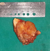
Then, the patient was referred to the Department of Oral Surgery for surgical treatment. The lesion was excised under general anesthesia, access to the mass was achieved via an intraoral approach, and the odontoma was elevated using a periosteal elevator. The capsule of the lesion was curetted. The odontoma appeared as a single mass. The excised specimen weighed 43.5 g and was rough and stony hard in terms of consistency. The first and the second premolars were extracted followed by suturing.
After excision, the specimen was sent for histopathological examination, which revealed conglomerate masses of dental hard and soft tissues arranged haphazardly (Fig. 5). Relatively large quantities of irregularly deposited mature tubular dentin, enclosing clefts and hollow circular spaces representing enamel-like tissue, were seen, which confirmed the diagnosis of a complex odontoma (Fig. 6). The patient has been kept under follow-up after the surgical excision and has had a healthy and disease-free life since the surgery.
Complex odontomas are slow-growing, expanding, and painless lesions. In rare cases, complex odontomas are associated with pain and inflammation. The relative frequency of complex odontomas varies between 5% and 30% among odontogenic tumors. Although they are common odontogenic lesions, they are rare compared to other variants. Complex odontomas occur at any age, but most of the reported cases have occurred before the age of 30 with a peak in the second decade of life as in the present case. Males are affected more than females with the ratio of 1.5 : 1 to 1.6 : 1. Further, most of the complex odontomas are located in the posterior mandible; the second most common site is the anterior maxilla.3,4 Some peripheral complex odontomas have been reported, such as those in the maxillary sinus.7
The etiology of complex odontomas is not clearly understood. However, several researchers have reported various etiological factors, including local trauma, infection, family history, and genetic mutation. They have also suggested that complex odontomas are inherited from a mutant gene or interference, possibly postnatal, with the genetic control of tooth development.8
Complex odontomas are usually asymptomatic and are associated with changes such as malformation, impaction, delayed eruption, malposition, cyst formation, displacement, resorption of the adjacent teeth, and expansion of the cortical plate. Symptoms that may be present include numbness in the lower lip, frontal headaches, swelling in the affected areas, and facial asymmetry.3,9 Pain is a rare symptom. In the present case, the patient presented with swelling and an expansion of the buccal cortical plate, which resulted in facial asymmetry.
Complex odontomas rarely erupt in the oral cavity; their eruption is different from tooth eruption: As the periodontal ligament is missing in the case of a complex odontoma, without the contractility of fibroblasts, an odontoma cannot erupt. Its increasing size may lead to sequestration of the overlying bone and hence, its eruption. Another reason could be the remodeling of jaw bones; for this, a dental follicle is required as it provides conductance and chemo-attraction for the osteoclast required for the eruption. Erupted complex odontomas are most often seen in the older population, but eruption at a younger age could be caused by bone remodeling that occurs due to the presence of dental follicles.2
Complex odontomas have been associated with inflammatory, infectious processes and hereditary anomalies (Gardner's syndrome and Hermann's syndrome). Their genetic inheritance through a mutant gene or interference has been suggested by Hitchin.8 Further, complex odontomas associated with pigmentation have been reported in the literature.10
The radiological appearance of complex odontomas depends on their development stage and degree of mineralization. The first stage is characterized by radiolucency due to a lack of calcification. Partial calcification is observed in the intermediate stage, while in the third stage, the lesion usually appears radiopaque with amorphous masses of the dental hard tissue surrounded by a thin radiolucent zone corresponding to the connective capsule histologically.11
Since our case was radiopaque with amorphous masses of the dental hard tissue and the radiolucent zone surrounding the lesion, the present lesion was considered to be completely mature and in the third stage. The absence of cortication at the superior aspect of the mass in the radiograph shows the eruption of the mass in the oral cavity.
In our case study, complex odontomas had to be differentiated from cemento-ossifying fibromas by the odontomas' tendency to associate with unerupted molar teeth and their property of being more radiopaque than fibromas.12 Periapical cemento-osseous dysplasia/focal cemento-osseous dysplasia (PCOD/FCOD) are mature fibro-osseous lesions, which may be solitary with dense radiopacities and a radiolucent rim; they are the entities most frequently confused with mature complex odontomas, usually the PCOD or FCOD form, in persons aged over 30 years, whereas a complex odontoma develops in much younger patients. PCOD is situated deep in the alveolar bone, whereas a complex odontoma often extends high into the alveolus towards the crest of the ridge.13
An ameloblastic fibro-odontoma has a greater soft tissue component than an odontoma; even when the amount of the hard tissue component increases, the complex odontoma has one mass of disorganized tissue in the center, whereas the ameloblastic fibro-odontoma has multiple scattered mature pieces of dental hard tissues. Odontoameloblastoma is an extremely rare condition and involves the simultaneous occurrence of an ameloblastoma and a complex odontoma.12
In our case, the lesion was found in the right mandibular posterior region, in which the second premolar and all molars were missing clinically. Radiolucencies observed in the lesion may relate to the pulp tissue histologically.14 Further, we considered the lesion to have been in the third stage, as it was completely radiopaque, with no sign of cortication observed in the superior periphery of the lesion.
The present case is interesting because of the unusually large size of the lesion involving all the posterior teeth of the mandibular region (right side) and full eruption in the oral cavity as most of the reported odontomas are intrabony lesions, and in exceptional cases, the odontoma erupts in the oral cavity.15 Thus far, to the best of our knowledge, the secondary effects of such large erupted odontomas have not been reported in the literature. In the present case, an ulcer was present on the adjacent surface of the buccal mucosa. This may have been present because of chronic irritation caused by the hard rough surface of the lesion, which was in contact with the mucosa.
Histologically, a complex odontoma consists primarily of a disordered mixture of odontogenic tissues, often spherical in shape. Cementum-like substances are often admixed with dentinoid structures. Small spaces with pulp tissue, enamel matrix, and epithelial remnants may be observed within the calcified mineralized masses of dentine of different qualities. A thin fibrous capsule, or occasionally, a cyst wall, is also seen surrounding the lesion. The present case revealed similar histological findings.
Treatment of complex odontomas is conservative surgical excision. The present lesion was also treated by using this method under general anesthesia. Holes were made on the thick mandible buccal cortical plate so as to intentionally fracture it and to reduce the expansion of the mandible and create facial symmetry.
Complex odontomas are tumors of odontogenic origin that may or may not be associated with systemic diseases. These are asymptomatic and are found on routine radiographs. They rarely erupt into the mouth and tend to be associated with impacted teeth. Despite their benign nature, their eruption into the oral cavity can give rise to pain, inflammation, infection, and ulceration, but large odontomas can cause cortical expansion facial asymmetry and traumatic ulcers. Therefore, it is important to diagnose these lesions as soon as possible and treat them appropriately so as to avoid complications.
Figures and Tables
Fig. 3
Panoramic radiograph shows the lesion as a well-defined radiopacity surrounded by a radiolucent halo with secondary inferior displacement and oblique horizontal impaction of the right mandibular second premolar.

Fig. 4
A. Sagittal section of cone-beam computed tomography (CBCT) shows a dense homogeneous ground-glass radiopaque matrix with a horizontally aligned distinct molar-like tooth, adherent to the posteroinferior third of the mass. B. Axial section of CBCT shows the similar features of the sagittal section with a buccolingual expansion of the mandible. C. Coronal section of CBCT shows the similar features of the axial and sagittal sections with a buccolingual expansion and a tooth in the inferior portion of the lesion. Three-dimensional images of the lesion in the anteroposterior view (D) lateral view (E) submental view (F) show the expansion of cortical bones.

Acknowledgments
The authors wish to express their sincere gratitude to Dr. Devi Charan Shetty, HOD, Department of Oral Pathology and Microbiology, and Dr. Sanjeev Saxena, HOD, Department of Oral and Maxillofacial Surgery and their respective teams, ITS Dental College, Murad Nagar, for their help with this case report.
References
1. Reichart P, Philipsen HP. Odontogenic tumours and allied lesions. London: Quintessence Pub;2004. p. 141–147.
2. Vengal M, Arora H, Ghosh S, Pai KM. Large erupting complex odontoma: a case report. J Can Dent Assoc. 2007; 73:169–173.
3. Dua N, Kapila R, Trivedi A, Mahajan S, Gupta SD. An unusual case of erupted composite complex odontoma. J Dent Sci Res. 2011; 2:1–5.
4. Serra-Serra G, Berini-Aytes L, Gay-Escoda C. Erupted odontomas: a report of three cases and review of literature. Med Oral Patol Oral Cir Bucal. 2009; 14:E299–E303.
5. Garcia-Consuegra L, Junquera LM, Albertos JM, Rodriguez . Odontomas. A clinical-histological and retrospective epidemiological study of 46 cases. Med Oral. 2000; 5:367–372.
6. Mupparapu M, Singer SR, Rinaggio J. Complex odontoma of unusual size involving the maxillary sinus: report of a case and review of CT and histopathologic features. Quintessence Int. 2004; 35:641–645.
7. De Visscher JG, Güven O, Elias AG. Complex odontoma in the maxillary sinus. Report of 2 cases. Int J Oral Surg. 1982; 11:276–280.
10. Kaur GA, Sivapathasundharam B, Berkovitz BK, Radhakrishnan RA. An erupted odontoma associated with pigmentation: a histogenetic and histological perspective. Indian J Dent Res. 2012; 23:699.

11. Philipsen HP, Reichart PA. Classification of odontogenic tumours. A historical review. J Oral Pathol Med. 2006; 35:525–529.

12. White SC, Pharoah MJ. Oral radiology: principles and interpretation. 6th ed. St. Louis: Mosby Elsevier;2010. p. 380–383.
13. Wood NK, Goaz PW. Differential diagnosis of oral maxillofacial lesions. 5th ed. St. Louis: Mosby;2007. p. 492.


 PDF
PDF ePub
ePub Citation
 Print
Print


 XML Download