

 PDF
PDF ePub
ePub Citation
 Print
Print


Introduction
Despite the development of several modern imaging modalities, the radiograph remains the first and the most important mode of investigation for evaluating jaw lesions.1 Many jaw lesions resemble each other radiographically, making them difficult to diagnose correctly.2 Focusing on the patient's history, along with an analytical approach to radiographs, helps to narrow the differential diagnosis.1 Some authors have developed classifications and guidelines to help clinicians evaluate radiographs more precisely.123 Wood and Goaz4 presented one of the most comprehensive classifications in 1997. In this classification, jaw lesions are categorized into three major groups depending on whether their radiographic appearance is completely radiolucent, mixed, or totally radiopaque. Each group is divided into several subgroups that represent a subclassification of the lesions based on more subtle radiographic differences. The main categories of their classification are periapical radiolucencies, pericoronal radiolucencies, inter-radicular radiolucencies, solitary cyst-like radiolucencies not necessarily contacting teeth, multilocular radiolucencies, solitary radiolucencies with ragged and poorly defined borders, multiple separate well-defined radiolucencies, mixed radiolucent-radiopaque lesions associated with or without teeth, periapical radiopacities, solitary radiopacities not necessarily contacting teeth, multiple separate radiopacities, and generalized radiopacities.
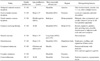
Based on a review of the literature, we propose a new subcategory, jaw lesions with a radiolucent rim, which contains eight entities (Table 1). When a clinician encounters a lesion with a radiolucent rim, he/she should first consider these entities in the differential diagnosis. This will help dental practitioners make more accurate diagnoses and better treatment plans based on patients' radiographs.
Literature review
We used general search engines and specialized databases such as Google Scholar, PubMed, PubMed Central, MedLine Plus, Science Direct, Scopus, and well-recognized textbooks to find relevant studies by using keywords such as "jaw disease," "jaw lesions," "radiolucent rim," "radiolucent border," and "radiolucent halo." More than 200 articles were found, of which 70 were broadly relevant to the topic. We ultimately included 50 articles that were closely related to the topic of interest. When the relevant data were compiled, the following eight lesions were identified as having a radiolucent rim: periapical cemento-osseous dysplasia, focal cemento-osseous dysplasia, florid cemento-osseous dysplasia, cemento-ossifying fibroma, osteoid osteoma, osteoblastoma, odontoma, and cementoblastoma. We propose a novel subcategory, jaw lesions with a radiolucent rim, which includes eight entities. The implementation of this new category can help improve the diagnoses that dental practitioners make based on patients' radiographs.
Osseous dysplasia/cemento-osseous dysplasia
The most common fibro-osseous lesion is cementoosseous dysplasia. It is considered to be a non-neoplastic fibro-osseous lesion in which bone is replaced by fibrous connective tissue along with cemental masses.5 In a 2005 publication, the World Health Organization described three clinical presentations of osseous dysplasia/cementoosseous dysplasia: periapical, focal, and florid osseous dysplasia/cemento-osseous dysplasia.6
In many cases of cemento-osseous dysplasias, evaluating intraoral radiographs suffices for diagnosis. The radiographic findings of all subtypes of cemento-osseous dysplasia lesions can be divided into an early stage (radiolucency without radiopaque inclusion), a mixed stage (radiolucency with radiopaque inclusion), and a mature (radiopaque) stage.7
Kawai et al.8 classified cemento-osseous dysplasia lesions into six types based on the following radiographic parameters: (A) radiolucency, (B) calcifications, (C) target appearance, (D) lobular or spherical calcified masses, (E) multiple periapical radiopacities, and (F) active hypercementosis in multiplex form. They studied the radiographic findings of 54 patients, and found that Type D was the most frequent pattern, manifesting as lobular or spherical calcified masses enveloped by a complete or partial radiolucent rim. In partially radiolucent rims, calcified masses were in close proximity to the peripheral bone without any association with the neighboring teeth. Type C was the second most common pattern, occurring in 35% of cases. The pathognomonic radiographic features in the diagnosis of cemento-osseous dysplasia lesions are features C and D. The presence of a radiolucent rim at the periphery of radiopacities is also very significant for diagnosis.8
Alsufyani and Lam9 found that the frequency of a radiolucent rim or band at the periphery of cemento-osseous dysplasia was 78.4% when the images were interpreted by oromaxillofacial radiologists, but 64% when analyzed by general dentists. Likewise, the studies of Alsufyani and Lam,7 Kawai et al.,8 Beylouni et al.,10 and Brannon and Fowler11 noted the presence of a complete or partial radiolucent rim in cases of cemento-osseous dysplasia.
Periapical cemento-osseous dysplasia
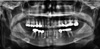
Periapical cemento-osseous dysplasia arises in the anterior mandible, generally in patients over 30 years of age. A female predilection has been found, and almost 70% of cases occur in blacks. Periapical cemento-osseous dysplasia has no symptoms and the teeth in the affected area remain vital. Hence, the lesion is detected in routine radiographs. A majority of periapical cemento-osseous dysplasias are delineated with a well-defined radiolucent border.12 The incidence of periapical cemento-osseous dysplasia is 2-3/1000 in the general population.13 The radiographic appearance of periapical cemento-osseous dysplasia may vary widely. In the early stage, lesions show a periapical radiolucent defect that subsequently develops very small radiopacities. In the later stage, tiny radiopaque foci enlarge, coalesce, and undergo further substantial opacification. They consist of dense cementum-like and/or ground-glass areas rimmed by a radiolucent halo.7 Eskandarloo and Yousefi12 reported a case in which a radiolucent rim was found around the radiopaque lesion in the teeth apices (Fig. 1). The same finding was reported by Komabayashi and Zhu.14
Focal cemento-osseous dysplasia
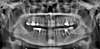
Focal cemento-osseous dysplasia was first described by Summerlin and Tomich15 based on the location of the involved bone (tooth-bearing areas of the posterior jaws and extraction sites). Although the etiology is unknown, some triggering factors such as trauma, caries, periodontal disease, infection, and systemic diseases have been identified. The periodontal ligament has been suggested as a probable origin for focal cemento-osseous dysplasia. Zegarelli et al.13 suggested that hormonal imbalance may also be a causative factor. It causes no symptoms and is detected only on radiographic examination. The majority of focal cemento-osseous dysplasia lesions occur in the mandible. Almost one third of patients with focal cementoosseous dysplasia present with local jaw expansion and mild discomfort.16 This entity has three stages of maturation: early (a well-defined radiolucency at the apices of the mandibular teeth), intermediate (a radiolucent-opaque lesion with a well-defined radiolucent halo) and late (a radiopaque lesion, often with an ill-defined periphery).17 Rao et al.18 described focal cemento-osseous dysplasia as a small well-defined radiopacity surrounded by a uniform radiolucent halo (Fig. 2).
Florid cemento-osseous dysplasia
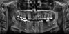
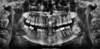
Florid cemento-osseous dysplasia has also been described using the terms multiple cemento-ossifying fibroma, sclerosing osteomyelitis, multiple osteoma, sclerosing osteitis, periapical cementoblastoma, multiple enostosis, gigantiform cementoma, and florid osseous dysplasia.1019 The exact etiology of florid cemento-osseous dysplasia is still unknown. Most authorities have suggested that the pathogenesis of florid cemento-osseous dysplasia is related to the periodontal ligament, because most florid cementoosseous dysplasias are seen close to the periodontal ligament with identical histopathologic features. However, a few authors have suggested that remnants of cementum in the bone after extraction might play a role in the etiology of this type of lesion.19 Florid cemento-osseous dysplasia may be familial, with an autosomal dominant inheritance pattern, but few examples of a familial pattern have been reported in the literature.20 It predominantly occurs in middle-aged to older black women, and the male-to-female ratio has been reported as 1 : 2.6.19 Florid cemento-osseous dysplasia most commonly appear as bilateral, symmetrical, and extensive lesions in all four posterior quadrants of the jaws, in the molar and premolar regions.10 Florid cementoosseous dysplasia has three types of radiographic appearance depending on its stage: the first or osteolytic stage (a well-defined radiolucent area with loss of lamina dura and periodontal ligament), the second or cementoblastic stage (small radiopacities appear in the radiolucent area due to the deposition of cementum-like droplets in fibrous tissue), and the last stage (lobular radiopacity throughout the lesion, surrounded by a radiolucent area).19 Beylouni et al.10 noticed cyst-like radiolucent areas around some radiopaque lesions in florid cemento-osseous dysplasia. Köse et al.19 and Kutluay Köklü et al.20 reported ovoid radiopaque masses in wide radiolucent spaces in the periapical areas of all molars in both quadrants of the mandible in the panoramic view (Fig. 3).
Cemento-ossifying fibroma
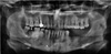
According to Wood and Goaz,4 this entity has also been categorized as a fibro-osseous lesion of the jaw arising from the progenitor cells of periodontium, which are able to produce fibrous tissue, cementum, bone or a combination thereof.421 This lesion has also been designated as central ossifying fibroma, cementifying fibroma, and ossifying fibroma.2122 The pathogenesis of cemento-ossifying fibroma is not clear, but trauma and congenital abnormalities in the maturation of dental tissue may play a key role in its development. It usually affects patients in their second to fourth decade of life, with a 1 : 5 male-to-female ratio. This lesion occurs much more frequently in the mandible (93%) than in the maxilla (7%), and it is most frequently seen in the molar region (61%), followed by the premolar (28%), and canine/incisor (11%) regions.212223 Clinically, it is a round, expansive, painless, slow-growing mass that may displace the roots of the adjacent teeth, sometimes causing root resorption.22 Radiographically, early-stage lesions are small and radiolucent. As they mature, they become mixed radiolucent and radiopaque lesions, and eventually become radiopaque.212223 K et al.,21 Chang et al.,22 Trijolet et al.,23 and Khan et al.24 have reported that the presence of a radiolucent rim around a mixed or radiopaque cemento-ossifying fibroma is a characteristic feature of this entity (Fig. 4).
Osteoid osteoma
Osteoid osteoma is a benign lesion that comprises 3% of all primary bone tumors and approximately 10% of bone tumors. Approximately 80% of lesions occur in the long bones, while less than 1% occur in the jaws; it is therefore a rare phenomenon in the orofacial region and might easily be misdiagnosed.25 Osteoid osteoma is most frequently observed in the second or third decades of life, and the average age at presentation is 19 years. It is more prevalent in females, with a male-to-female ratio of 1 : 2. Most patients present with nocturnal pain of the involved bone that is usually alleviated by non-steroidal anti-inflammatory drugs.26
An et al.26 found that approximately 80% of cases had a mixed or high-density appearance. More than 50% of the cases were accompanied by surrounding sclerosis and 24% presented with periosteal reaction. The presence of a radiolucent rim or border around mixed or radiopaque osteoid osteoma was reported by Ida et al.,25 An et al.,26 and Liu et al.27
Osteoblastoma
Osteoblastoma is a benign and slow-growing bone neoplasm characterized by the formation of osteoid and bone with a great number of osteoblasts. This lesion comprises up to 1% of all primary bone tumors and 15% of maxillofacial lesions.28 It has been found to occur in patients ranging in age from three to 78 years, with a mean age of 23 years. Patients between 20 and 30 are most commonly affected. Men are affected twice as frequently as women. Instances of mandibular involvement have been found to outnumber cases of maxillary involvement by 74% to 26%, and the mandibular body is most commonly affected.2829 The primary etiology of osteoblastoma is not clear, but trauma, inflammation, an abnormal local response of tissue to injury, and local alterations in bone physiology have been documented as possible causative factors.30 The clinical features of osteoblastoma include mild pain, swelling, and expansion of the cortical bone. It is characterized by a limited growth pattern and does not exceed 4 cm in diameter.30 The pain does not have a nocturnal pattern, and is not relieved by aspirin or other non-steroidal anti-inflammatory drugs, as is the case for osteoid osteoma.31 A combination of radiopaque and radiolucent patterns, depending on the degree of calcification and the absence of a perilesional sclerotic border, is a common radiographic feature for osteoblastoma.30 Jones et al.31 found that more than half of the cases analyzed in their study had mixed or complete radiopaque patterns. Manjunatha et al.28 reported similar findings.
Odontoma
Odontoma is the most frequent benign odontogenic tumor, and involves a combination of mesenchymal and epithelial dental elements. A variety of dental tissues, including enamel, dentin, cementum, and, in some cases, pulp tissue, can be found within the tumor. Two kinds of odontoma have been identified according to the latest classification of the World Health Organization (2005)6: complex odontoma and compound odontoma, which is twice as common as the complex type. Odontoma manifests in three clinical forms: central (intra-osseous), peripheral (extra-osseous or soft-tissue), and erupted odontoma.34 The site that is most commonly affected by compound odontoma is the anterior portion of the maxilla, where it appears as a pericoronal lesion around unerupted teeth or as an inter-radicular lesion. Odontoma is usually unilocular and contains several radiopaque, small denticles (tooth-like structures).34 Complex odontoma grows slowly and non-aggressively, and is slightly more common in the mandible, especially in the second and third molar areas.3435 The etiology of odontoma is unknown, but some researchers have suggested that trauma, infection, or genetic mutations may be causative factors.36 Odontomas are usually slow-growing and nonaggressive, and occur most frequently in children and adolescents.37 Odontoma does not have a marked gender predilection. Clinically, odontomas are asymptomatic lesions that are often associated with altered tooth eruption in the permanent or deciduous dentition. They are usually diagnosed based on routine panoramic and/or intraoral X-rays, or by assessing the cause of delayed tooth eruption. It is believed that 37%-78% of all eruption disorders in the dentition are caused by odontomas.37 Occasionally, odontomas are discovered due to the presence of retained deciduous teeth, eruption problems, pain, swelling, infection or inflammation, or dental malposition.353637
Radiographically, compound odontomas present as welldelineated lesions containing radiodense foci with a radiolucent halo. Several follicular structures contain denticles, which are separated by fibrous septae. However, in complex odontomas, opaque foci present as haphazard masses with no similarity to dental structures.38 The presence of a radiolucent rim on the border of odontoma has been noted by Piattelli et al.,35 Reddy et al.,36 Hidalgo-Sànchez et al.,38 Philipsen et al.,39 and Garcia-Consuegral et al.40 (Fig. 5).
Cementoblastoma
Cementoblastoma constitutes less than 6% of all odontogenic tumors. According to current World Health Organization classification of odontogenic tumors, it is classified as an ectomesenchymal tumor with or without odontogenic epithelium.41 Cementifying fibroma and cementoblastoma are considered to be true neoplasms of cemental origin.414243 Cementoblastoma originates from the roots of teeth, and is marked by the formation of cementum-like tissue.4445 Clinically, the lesion appears as a nodular expansive mass in the alveolar ridge area that is hard to elastic in consistency.45 The tumors have been reported to range in size from 0.5 cm to 5.5 cm, with a mean diameter of 2.1 cm.46 Cementoblastoma is usually asymptomatic and is generally detected upon radiographic examination.4243 Symptoms may be completely absent, but if present, pain, swelling, and cortical expansion are frequent findings.424748 Paresthesia of the lower lip or pathologic fracture of the jaws rarely occurs.42434445464748 It can also act in a locally aggressive manner, resulting in bony expansion, root resorption, displacement of adjacent teeth, and jaw deformity. Jaw expansion and perforation of the cortex are common findings in recurrent tumors.42 Most benign cementoblastomas occur in the mandible.474849 The mandible is affected three times as often as the maxilla. The most commonly affected tooth is the mandibular first molar, which remains vital.4748 Benign cementoblastoma has also been reported in the maxillary sinus, and is accompanied by deciduous and unerupted permanent teeth.414345 Caucasians are more commonly affected than blacks. The male-to-female ratio of its prevalence is 2.1 : 1. Young adults in their second and third decades of life are most commonly affected.4142434445464748
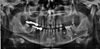
Radiographically, cementoblastoma presents as a welldefined radiopaque mass surrounded by a thin, radiolucent rim of non-mineralized tissue, in close association with roots of the involved tooth (Fig. 6). Root resorption, loss of usual outline, and obliteration of the periodontal ligament are common radiographic features.5051 Attachment of the lesion to the roots of the involved tooth is nearly pathognomonic. Additional radiographic findings consist of invasion of the root canal, bony expansion, displacement and involvement of adjacent teeth, and cortical erosion.29 A radiolucent rim at the periphery of cementoblastoma has been pointed out by Sharma,41 Sankari and Ramakrishnan, 42 Kumar et al.,43 Iannaci et al.,45 Huber and Folk,47 Bilodeau et al.,48 Harada et al.,49 Pacifici et al.,50 and Monks et al.51
Discussion
We propose the novel subcategory of jaw lesions with a radiolucent rim, which has the advantage of helping clinicians to rule out improbable diagnoses and arrive at the correct diagnosis timely and in a logical manner. This subcategory consists of eight entities: periapical cementoosseous dysplasia, focal cemento-osseous dysplasia, florid cemento-osseous dysplasia, cemento-ossifying fibroma, osteoid osteoma, osteoblastoma, odontoma, and cementoblastoma.
Of particular note is the pattern of female predominance in all of the above lesions except for cementoblastoma. With respect to location, only compound odontoma has a tendency to occur in the maxilla, and the remaining lesions are most commonly encountered in the mandible. However, florid cemento-osseous dysplasia can involve both jaws simultaneously. Moreover, with the exception of compound odontoma and periapical cemento-osseous dysplasia, the posterior mandible is the most common site of involvement. Osteoid osteoma and osteoblastoma have been reported to cause pain, while the other six jaw lesions in this category remain asymptomatic until they are noticed due to cortical expansion or during a routine examination. Most jaw lesions a with radiolucent rim manifest as periapical lesions with or without association with the teeth, but odontomas occur as pericoronal lesions. However, the ability to expand the cortical plates varies among these lesions; some entities, such as periapical cementoosseous dysplasia, remain less than 1 cm in diameter with no expansion, while cemento-ossifying fibroma and cementoblastoma are capable of expanding the jaw bone considerably.41012161719222628293445
This newly-proposed category of jaw lesions allows practitioners to narrow the differential diagnosis and arrive at the most probable diagnosis via a systematic method.
 XML Download