

 PDF
PDF ePub
ePub Citation
 Print
Print


Introduction
Recent technological advances have significantly impacted the practice of dentistry. Technological innovations are continuing to offer innovative solutions to a variety of traditional challenges in routine clinical dental practice. The current surge in technological innovation has led to the emergence of a large number of applications that contribute to the delivery of superior-quality health care. Radiology is an extremely technology-driven field, and new imaging modalities are constantly developed in order to provide more efficient and effective services to patients.
A major advance in recent years has been the development of tablet computers. With high-resolution displays and touch-screen interfaces, tablet computers are becoming an integral part of many industries, including the healthcare industry. Tablet computers can be used for multiple clinical applications, such as viewing radiographs and accessing a patient's electronic health records - whether they are located on a local storage device or on a cloud server - and can help in scheduling, consultation, and patient education. Additionally, information can be accessed and shared more efficiently using tablet computers, eliminating the need for extensive paper records.1
Tablet computers appear to be able to offer a robust and mobile solution for displaying images, providing quick access to a patient's medical records and allowing easy access to patient educational material. However, before they can be fully integrated into patient care, these new and evolving display devices must be tested to ensure that they do not compromise interpretative accuracy. A review of the literature reveals a small but growing number of scientific articles evaluating the performance of tablet computers in viewing radiographic images.234 However, most of these studies have dealt with medical radiography, not dentistry. John et al.5 evaluated the representation of common emergency conditions in computed tomographic (CT) and magnetic resonance images, finding that the diagnoses made using tablet computers were in agreement with those made on picture archiving communication system (PACS) workstations. Johnson et al.6 found that pulmonary embolism was accurately identified in CT images viewed on an iPad, and that the diagnoses made using an iPad were equivalent to those made using PACS workstations. In the field of dentistry, Shintaku et al.7 found no significant differences between a tablet computer (the iPad 2) and an LCD monitor in the detection of interproximal caries in digital intraoral radiographs.
In light of the growing popularity of these portable devices and the lack of an adequate number of studies exploring the dental applications thereof, we determined that evaluating the accuracy of tablet computers in the identification of fundamental anatomical landmarks would provide a baseline assessment that appears to be absent in the current literature. The purpose of our study was to evaluate and compare the diagnostic visibility of anatomical landmarks in panoramic and lateral cephalometric radiographs on a standard medical grade PACS monitor and on an iPad 5.
Materials and Methods
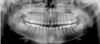
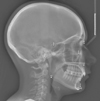
An existing set of 1000 digital radiographs (500 panoramic radiographs and 500 lateral cephalometric radiographs) were retrieved from the archive of the Section of Oral and Maxillofacial Radiology of the University of Connecticut School of Dental Medicine. The images were taken between the years of 2005 and 2012 using conventional radiography techniques, and were acquired with an Orthoralix 9200 machine (Gendex, Norcross, GA, USA). The exposure parameters were 75 kVp, 5 mA, and 12 seconds for the panoramic radiographs and 2.5 seconds for the lateral cephalometric radiographs. The images were then scanned into a computer and saved as JPEG files with unadjusted exposure levels. Therefore, the observers were not allowed to manipulate the density and contrast of the images. The images were de-identified to exclude each patient's name, age, and gender, and were viewed in Microsoft Office PowerPoint 2010 (Microsoft, Redmond, WA, USA). The images were viewed on an LCD monitor using an HP Compaq LA2205wg desktop PC (96 DPI, 1680×1050 pixels, HP, Palo Alto, CA, USA) and on an iPad Air (fifth generation; height, 240 mm, width, 169.5 mm; diagonal dimensions, 9.7 inches; Cupertino, CA, USA) with a Retina display (2048×1536 pixels, 264 ppi). In the panoramic images, the following landmarks were identified: the sigmoid notch, mandibular condyle, mandibular ramus, angle of the mandible, inferior border of the mandible, coronoid process, mental foramen, glenoid fossa, pterygomaxillary fissure, floor of the maxillary sinus, anterior nasal spine, nasal fossa, external oblique ridge, alveolar crest, articular eminence, and zygomatic arch (Fig. 1). In the lateral cephalometric radiographs, the following landmarks were identified: the porion, orbitale, molars, incisors, sella, nasion, pogonion, gnathion, menton, gonion, point A, and point B (Fig. 2). Two examiners (one senior radiology resident and one senior dental student) reviewed the images on both displays independently under dim ambient light. The examiners initially reviewed ten panoramic radiographs and ten lateral cephalometric radiographs to verify interoperator agreement in landmark identification. The following scoring system was then used to rate the visibility of each landmark on the two displays: 0, the landmark was not visualized on either of the displays; 1, the landmark was visualized on only one display; 2, the landmark was visualized on both displays with a level of clarity appropriate for diagnostic use; 3, the landmark was visualized on both displays, but especially clearly on one or the other.
The images were also rated based on their level of exposure, and independently classified by each reviewer as normal, overexposed, or underexposed. An image was considered underexposed if it was too light and the differences between anatomical structures were not clearly detectable. An image was classified as overexposed if it was too dark and the differences between anatomical structures were not clearly detectable. Since the images were acquired using conventional techniques, scanned directly into the computer, and saved as JPEG images, no image processing or adjustment of density and contrast was allowed. Any disagreement was resolved by referring to a senior oral and maxillofacial radiologist. The data were entered into Microsoft Office Excel 2010 (Microsoft, Redmond, WA, USA) and subjected to statistical analysis using Pearson's correlation test. Cohen's kappa analysis was performed to verify interoperator reliability.
Results
A total of 1230 images were retrieved from the archive, of which 730 were discarded because they did not have both a preoperative panoramic image and a lateral cephalometric image or because they were of poor quality. Of the 500 cephalometric images selected, 30 were underexposed and 470 were normal. Of the 500 panoramic radiographs, 10 were underexposed, 24 were overexposed, and 466 were normal. Cohen's kappa for interoperator reliability was 0.85. The visibility of landmarks on the standard monitor and the iPad was compared using the Student's t-test. The statistical analysis failed to show any significant difference between the visibility and clarity of the landmarks in either the panoramic radiographs or the cephalometric radiographs. However, the iPad was able to provide better clarity and visibility for certain landmarks (the anterior nasal spine, the external oblique ridge, and the mental foramen in the panoramic radiographs, and the porion, the orbitale, and the molars in the cephalometric radiographs) (Tables 1, 2, 3, 4, 5). The score of 1 was eliminated from the analysis, since none of the landmarks were assigned the score of 1. Pearson's correlation test found a statistically significant correlation between the ability of the iPad to show the mental foramen and the level of exposure (P<0.05). The iPad showed the mental foramen more clearly in underexposed images. Pearson's correlation test also found a statistically significant correlation between the clarity of the porion, the orbitale, and the molars in cephalometric radiographs and the level of exposure (P<0.05). The iPad was able to render the above landmarks more clearly in underexposed radiographs.
Discussion
The evolution of mobile device technology has dramatically changed how images are viewed in the domain of consumer electronics. Tablet computers are quickly becoming ubiquitous. Their very high screen resolution and processing capabilities can be used in routine dental practice for visualizing images and viewing electronic health records. The biggest advantage to end users is that these devices are lightweight and mobile. While these devices are extensively used in other industries, their application in dentistry has remained limited.
Traditionally, LCD and CRT monitors have been used for radiographic image evaluation.8910 A very limited number of studies have found that the accuracy of caries detection in intraoral images was not affected by the use of a consumer-grade or medical display system. Shintaku et al.7 evaluated the use of an iPad for diagnostic tasks, such as the detection of caries in intraoral images, and found that tablet computers are very reliable for the detection of caries. A more recent study by Kallio-Pulkkinen et al.11 studied caries and apical pathology in panoramic radiographs viewed on tablet devices under dim light and suboptimal conditions, and found tablet computers to be effective for the detection of pathology. To the best of our knowledge, no other study has evaluated panoramic radiographs viewed on tablet computers, which indicates that the use of tablet computers to evaluate panoramic and cephalometric images has not been adequately studied.
A definite gap in knowledge exists regarding the fundamental issue of anatomical landmark identification on panoramic and cephalometric images displayed on tablet devices. In this study, we evaluated 1000 radiographs, including 500 panoramic and 500 cephalometric images. The visualization of anatomical landmarks is essential for image analysis and subsequent diagnosis. The adequate visualization of anatomy is key for the diagnosis of pathology associated with a given area. In our study, we found that a tablet computer was able to display the anatomical landmarks adequately, in a manner completely comparable to a traditional medical-grade PACS monitor display system. Some of the images that were ultimately included in our analysis were underexposed and overexposed, but the level of exposure did not affect the identification of the landmarks. While the PACS monitor allowed the images to be magnified using a mouse, the tablet device was easier to navigate due to its touch-screen capability. The ability to magnify or move the image using the pinch-screen function of the touch-screen interface was reported by the evaluators to have been very convenient.
Hellen-Halme et al.121314 found that when ambient light was <50 lx, as recommended by the American Association of Physicists in Medicine,15 no differences in the diagnostic accuracy of proximal caries lesions were observed among well-adjusted standard color displays, Digital Imaging and Communication in Medicine (DICOM) pre-calibrated color displays, and monochromatic displays. In our study, we standardized the ambient lighting and noise levels and we used the same settings during image evaluation with both display systems and both types of images.
In routine panoramic image evaluation, at least 20 anatomical landmarks are typically studied as part of the radiographic survey,8 and we simulated this process by evaluating the ability of the operator to use both displays to adequately visualize these landmarks. For cephalometric image evaluation, we evaluated all of the cephalometric landmarks that are commonly used in orthodontic analyses. We evaluated the ability of the operators to use both displays to identify these landmarks, finding no statistically significant differences associated with either the imaging display or the operators in the evaluation of these landmarks. However, some anatomical landmarks, such as the mental foramen, the anterior nasal spine, and the external oblique ridge, were visualized more clearly on the tablet than on the PACS monitor, especially in underexposed images, but not to an extent that affected the ability to identify the anatomical landmark in question. In cephalometric image analyses, certain anatomical landmarks, such as the porion, the orbitale, and the molars, were far more easily identified and clearly visualized on the tablet. The technical specifications of newer tablet systems are significantly better than those of average consumer-grade monitors but are unlikely to be better than those of high-end medical-grade display systems. In our study, although the technical specifications of the tablet systems were not significantly superior to those of the PACS monitor, the operators stated that the ability to easily magnify and manipulate the images on the touchscreen tablet systems may have significantly contributed to their finding that some landmarks were more clearly visualized on the tablets. Nevertheless, the ability to identify specific anatomical landmarks did not differ between the display systems.
While this study evaluated image display, several applications can be installed on a tablet computer to enhance patient care. Access to a patient's electronic health records and the ability to review radiographic images and educational images and videos, including potential treatment plans, could be especially valuable features. With their considerable advantage in terms of convenience and mobility, tablet devices could potentially eliminate the necessity for a workstation next to every single dental chair. This could be both a way to reduce costs and an economical and functional way of enhancing patient and provider interactions.
Dental applications, such as so-called 'orthodontic apps,' are being developed for tablet computers and smartphones, have become more reliable, and are being used with increasingly frequency in dental offices16. Future studies are essential in order to evaluate other applications of tablet computers, including their use with different radiographic imaging modalities and as a way of accessing electronic health record systems.
Tablet devices, such as the iPad, are comparable and at times superior in their ability to depict anatomical landmarks on panoramic and cephalometric radiographs. Due to features such as high resolution, touch-screen interface, long battery life, and light weight, these devices are highly portable and are a very good mobile alternative to conventional PACS monitor display systems for evaluating panoramic and cephalometric radiographs.


 XML Download