

 PDF
PDF ePub
ePub Citation
 Print
Print


Introduction
The detection of proximal dental caries is a challenge in dentistry. Most of these caries cannot be detected through optical observation,1 and radiographic examinations may be needed to detect these carious lesions. Bitewing radiography is the most common technique used for the detection of proximal dentinal caries, in addition to clinical examinations.2
In the last decade, intraoral digital radiographic systems have been introduced to dentistry, providing many advantages, such as lower radiation exposure, ease of digital storage and electronic transmission, and the absence of the requirement for darkroom equipment and wet processing.34 Moreover, digital radiographs can be modified using image processing software to improve the quality of the data and to reduce the factors that interfere with image quality.5 Furthermore, dentists can correct or adjust the contrast and brightness of the radiographs in ways suitable for specific diagnostic goals, such as the detection of carious lesions.67
The rapid emergence of digital imaging techniques in intraoral radiography necessitates recommendations about optimizing electronic image display settings for diagnostic purposes.8
A large number of software options and algorithms are available for digital radiography, with a range of purposes. Some of these features may affect the diagnostic accuracy of the radiographs through alterations to the contrast and brightness of images.
Post-processing algorithms are used in digital radiography to optimize the transfer of radiographic information to the observer. Conventional film radiographs can only be presented in the negative mode ('bones white'), whereas the grey-scale polarity of digital images can be inverted ('bones black'). Although several studies exist on grey-scale reversal in the field of general medicine with varying results,9101112 few such studies have been conducted in the field of dentistry.13 Haak et al.13 proposed a reversed grey-scale algorithm for detecting proximal dental caries, concluding that such a manipulation was not able to optimize the detection of proximal dental caries and aggravated the detectability of dentinal lesions. Due to a lack of sufficient published data, an obvious demand exists for more studies addressing this issue in order to arrive at a secure, evidence-based conclusion.
The present study was conducted to experimentally analyze the ability of the reverse contrast mode of intraoral digital radiography to detect proximal dentinal caries.
Materials and Methods
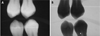
This comparative in vitro study included 80 maxillary and mandibular premolar teeth that were extracted for orthodontic reasons. The institutional review board and ethics committee of our university approved the protocol of this study (approval number 15004). The teeth were clinically analyzed, and were excluded if they had any restorations or visible proximal dentinal caries or cavities. The teeth were mounted in a straight line in order to avoid overlapping on the radiographs and embedded in plaster of Paris along with their roots. Digital radiographs were taken from the selected teeth using an intraoral radiographic device (Orix 65, Ardet, Milano, Italy) with a charge-coupled device sensor and an active area equivalent to 30×20 mm (Cygnus Media 3.0, Ritter, Asheville, NC, USA) using a paralleling technique. The pairs of blocks and teeth were fixed in a modified jig during radiography to maintain their precise geometry and ensure the repeatability of the radiographs. The exposure environment was set at 80 kVp, 8 mA, and 0.2 seconds. The digital radiographs were then displayed using the Cygnus Media 2002 software (Ritter, Asheville, NC, USA), both in the original form and using the reverse contrast mode, on a 17-inch SVGA monitor (Flatron 700B, LG Electronics, Seoul, Korea) with a resolution of 800×600. Figure 1 presents the digital radiographs in their original form and using reverse contrast mode. The radiographs were then examined by four experienced oral and maxillofacial radiologists with a mean experience of nine years (range, 5-20 years), and their views about the presence or lack of proximal dentinal caries were recorded on a five-point Likert scale from 0 to 4 (0, caries definitely are not present; 1, caries probably are not present; 2, suspicious; 3, caries are probably present; and 4, caries definitely are present). Disagreements were resolved by discussion and a final consensus was reached. No time limit was set for viewing the images.
After the radiography was performed, the teeth were cut in the mesiodistal direction using a diamond bur (Drendel+Zweiling, North Rhine, Germany) under a copious supply of water. The samples were kept in 10% formalin for one week. Thereafter, they were kept in a solution containing 700 cc of distilled water, 200 cc of formalin, and 100 cc of pure formic acid for 25-30 days. This solution was changed every three days. After decalcification of the teeth, they were taken out of the solution, washed with normal saline, and neutralized with lithium carbonate solution. After the tissue circulation process, paraffin blocks were prepared, from which 5-µm sections were prepared in the mesiodistal direction. Four chambers were prepared from each sample, stained with hematoxylin and eosin, and analyzed by a maxillofacial pathologist using an optic microscope (ALPHA PHOt 2, Ys-Nikon, Tokyo, Japan) at 10× and 40× magnification in order to confirm the presence or lack of proximal dentinal caries. Premolars with proximal caries were considered to be positive controls after undergoing the same treatment as the above samples, and the presence or lack of proximal dentinal caries was analyzed according to the standards presented in a previous study14 as well as by comparison with control samples. The chambers prepared from each sample with dentinal caries were considered to be positive samples. The premolars were categorized into two groups based on the radiologists' score assessing the presence or absence of proximal caries. Teeth with scores of 0-1 and 2-4 were considered caries-free and carious, respectively. After identifying the healthy and carious samples using each methods, a 2×2 table was used to compare them with the gold standard diagnostic technique. The sensitivity, specificity, positive predictive value, negative predictive value, and accuracy of both techniques were calculated based on generally accepted formulas.
The data were entered into SPSS version 21 (IBM Corp., Armonk, NY, USA). The chi-squared test was applied to compare the performance of the two methods on the above-mentioned indices. P-values <0.05 were considered to indicate statistical significance.
Results
Our statistical analysis found that the sensitivity of the original radiographic technique for proximal dentinal caries was 72.5% (95% confidence interval [CI]: 69%-76%). The sensitivity of the reverse contrast mode for the detection of proximal dentinal caries was 63.1% (95% CI: 59.7%-66.5%). The sensitivity of the original digital radiography technique was significantly higher than that of the reverse contrast mode (p<0.05).
The specificity of the original radiography technique for detecting proximal dentinal caries was 90% (95% CI: 87.7%-92.3%). The specificity of the reverse contrast mode for detecting proximal dentinal caries was 89.4% (95% CI: 87%-91.8%). No significant difference was found between the specificity of these two methods for the detection of proximal dentinal caries (p>0.05).
The positive predictive value of the original radiographic technique for detecting proximal dentinal caries was 87.2% (95% CI: 84.6%-89.8%). The positive predictive value of the reverse contrast mode for detecting proximal dentinal caries was 87.1% (95% CI: 84.5%-89.7%). No significant difference was found between the predictive positive value of these methods for proximal dentinal caries (p>0.05).
The negative predictive value of the original radiographic technique for proximal dentinal caries was 76.5% (95% CI, 73.2%-79.8%). The negative predictive value of the reverse contrast mode for proximal dentinal caries was 73.5% (95% CI, 70%-77%). No significant difference was observed between these two methods regarding the predictive negative value for proximal dentinal caries (p>0.05).
The accuracy of the original radiographic technique for detecting proximal dentinal caries was 80.9% (95% CI: 78.7%-83.1%). The accuracy of the reverse contrast mode for detecting proximal dentinal caries was 78.8% (95% CI: 75.5%-81.1%). No significant difference was found between these methods regarding the accuracy of detecting proximal dentinal caries (p>0.05) (Table 1).
Discussion
In the present study, the reverse contrast mode was found to have a lower sensitivity for the detection of proximal dentinal caries. No significant difference was observed between the traditional radiographic method and the reverse contrast mode regarding the specificity, positive predictive value, negative predictive value, and accuracy.
Previous research has conclusively demonstrated the superiority of digital radiography over conventional film-based radiography, especially regarding dentinal caries.1516 Thus, current research on digital radiography has generally progressed from comparing digital radiography with conventional film-based radiography to the investigation of how software and post-processing factors can affect the diagnostic reliability of digital radiography. Moreover, due to the lack of sufficient evidence regarding the reverse contrast algorithm for the detection of dentinal caries, it may be difficult to compare the results of our study with those of other studies.
Moystad et al.17 compared original digital radiographs and digitally enhanced radiographs with regard to the detectability of proximal dentinal caries. They found that both techniques had a similar diagnostic accuracy, corresponding to the results of the present study. Further, Tantanapornkul et al.18 found that the reverse gray-scale and contrast-brightness options did not lead to any significant difference in terms of the sensitivity and specificity in detecting proximal dentinal caries in comparison with original digital radiography, which was again mostly in agreement with the results of the present study.
Dove and McDavid19 evaluated the influence of the gray-scale reversal of enhanced bitewing images on the detection of proximal caries. They found that the digital processing techniques they employed did not enhance the diagnostic accuracy of dental images. Moreover, no significant differences were observed regarding diagnostic accuracy between non-enhanced digital images and conventional film-based images for the detection of proximal caries.
In a study comparable to ours, Haak et al.13 evaluated the effect of the gray-scale inversion display on the detection of approximal caries. They concluded that the gray-scale reversal of digital radiographs did not optimize the detection of approximal caries and aggravated the detectability of dentinal lesions.
Jorgenson et al.20 conducted a study comparing conventional film-based, original digital, and inversion-enhanced digital radiographs for the detection of interdental vertical bone defects. They found that the original digital radiographs were better than conventional film radiographs, and that the conventional film radiographs were better than inversion-enhanced digital radiographs in the detection of bone defects.
Verdonshott argued that image manipulation did not affect the sensitivity and specificity of images used to detect enamel caries, but did increase the sensitivity and decrease the specificity in the detection of dentinal caries.21 These divergent findings can be attributed to the type of sensor and the more accurate software and hardware used in the above study.
The present study had some limitations. For instance, soft tissue was not simulated in our study. Moreover, all of the teeth examined were premolar teeth. In order to overcome these limitations, similar studies may need to be carried out on human subjects. Another limitation was that the radiographic examination was performed using a predetermined exposure setting. Different exposure environments would probably have altered our findings.
In conclusion, the sensitivity of the original digital radiography technique for detecting proximal dentinal caries was significantly higher than that of the reverse contrast mode. However, no statistically significant differences were found regarding specificity, positive predictive value, negative predictive value, or accuracy. In general, the use of the reverse contrast mode in the detection of proximal dental caries should be regarded as an adjunct to other diagnostic methods, and not as an essential diagnostic aid. Due to the lack of sufficient studies dealing with this issue, more studies, especially clinical trials, are needed to validate our results.

 XML Download