

 PDF
PDF ePub
ePub Citation
 Print
Print


Introduction
Vertical root fracture is the most severe form of longitudinal tooth fracture,1 which can cause an inflammatory process, leading to bone resorption and the formation of granulation tissue.2 Clinical and radiographic signs of vertical root fractures are variable and nonspecific; they may be similar to those of periodontal lesions and endodontic failures.3,4,5 Nowadays, in the diagnosis of a vertical root fracture, the patient's history and symptoms, such as pain, swelling, the existence of deep isolated periodontal pockets, and the accompanying periapical and lateral radiolucencies associated with the root, provide valuable information.6 A radiographic diagnosis of vertical root fractures is difficult due to the various fracture patterns and the fractures are not found on the radiographs in many cases.7 Unfortunately, only one-third of these fractures can be diagnosed on conventional radiographs.8,9,10 Conventional radiography can show only two dimensions of the three-dimensional (3D) anatomical structures.11 Due to the limitations of intraoral radiographs, 3D imaging systems such as conventional computed tomography (CT), cone-beam computed tomography (CBCT), and multi-detector computed tomography have been introduced. In recent years, various studies have been performed to investigate the accuracy of each of these systems. According to various studies, CT systems are highly accurate in diagnosing root fractures.3,4,5,6,7,8,9,10,12,13,14,15 However, despite the many capabilities of CT, the main disadvantages of its dental applications are the high radiation dose in comparison with conventional radiography, the existence of artifacts, and the relatively low spatial resolution.13 These limitations led to the development of CBCT, which is considered a better alternative.3
The aims of this study were to investigate the accuracy of CBCT in detecting vertical root fractures and to assess the influence of root canal filling and intra-canal prefabricated posts on the visibility of a root fracture.
Materials and Methods
Ninety-six human mandibular premolars and molars without fracture, periapical pathology, root resorption, or any other anomaly were collected. The teeth had not undergone any restorative or root canal treatment. The teeth were inspected under a stereomicroscope (Zeiss Stemi SV6, Zeiss, Gottingen, Germany) to confirm the absence of vertical root fractures. Access cavity preparation and root canal treatment were done with a Pro Taper Rotary System (Dentsply Maillefer, Tulsa, OK, USA) up to size F3.
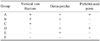
In order to create root fractures in half of the teeth (24 canals of premolars and 24 distal canals of the molars), the teeth were mounted in acrylic resin after covering the root surfaces with a thin layer of modeling wax to simulate soft tissue. Then, a screw driver-tape wedge was placed inside the canals, and fractures were created in the intended canals by applying gentle pressure on the hammer. Therefore, in 48 canals (half of the teeth), fractures were created, and well-fitting gutta-percha (Pro Taper F3) was placed in the root canal in 32 teeth (16 canals of premolars and 16 distal canals of molars); further, gold-plated screw posts (Nordin, Montreux, Switzerland) having the sizes of #4 and #5 were placed in the other 16 canals (8 canals of premolars and 8 distal canals of the molars). The same procedures were performed for the canals without fracture, and therefore, the teeth were divided into the six groups (8 premolars and 8 molars in each group) listed in Table 1.
The teeth were again evaluated using a stereo microscope to ensure the absence of vertical root fractures in the teeth without fracture. Then, the teeth were coded, and a wax rim was made from 3 to 4 layers of wax designed on the acrylic base in the shape of the mandible.
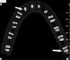
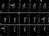
CBCT scans were obtained using the denture scan mode of a Newtom 5G system (QR s.r.l., Verona, Italy) set at 110 kV. Axial and multi-planar reformation (MRP) images were evaluated by using NNT viewer software version 3.0 (QR s.r.l., Verona, Italy). In this study, the slice thickness was 0.3 mm and the interval space was 0.3 mm. All of the obtained scans were examined by three oral and maxillofacial radiologists in the axial (Fig. 1) and MPR planes (Fig. 2) in a low-light room by using a Flatron 18.5-inch monitor (LG, Seoul, Korea). The observers were free to choose the magnification degree. All of the teeth were examined for the presence or absence of a vertical root fracture.
The data were analyzed by using IBM SPSS 20.0 statistical software (IBM Corp., Armonk, IL, USA). The sensitivity, specificity, positive predictive value (PPV), negative predictive value (NPV), and likelihood ratio (LR+, LR-) with 95% confidence interval were calculated based on an evaluation of at least two observers for each tooth. The kappa coefficient was used to assess the agreement among all the observers, and a p value of 0.05 was considered significant.
Results
Our results showed that the specificity in the diagnosis of a vertical root fracture using CBCT was 100% in all groups. Further, the sensitivity of vertical root fractures in the teeth with gutta-percha and without prefabricated posts was a little higher than in those without gutta-percha and prefabricated posts even though there was no statistical significance. The sensitivity in the diagnosis of vertical root fractures in the groups having teeth with gutta-percha and prefabricated posts was lower than in the other groups. The PPV was 100% in all of the groups, and the NPV in the groups containing teeth with gutta-percha and without prefabricated posts was higher than in the other groups; further, NPV in those without prefabricated posts and gutta-percha teeth was higher than in the groups containing teeth with gutta-percha and prefabricated posts.
In the case of the first observer, the kappa coefficient was 0.875±0.049 and the p value was <0.001; in the case of the second observer, the kappa coefficient was 0.896±0.045 and the p value was <0.001; and in the case of the third observer, the kappa coefficient was 0.771±0.064 and the p value was <0.001. The sensitivity, specificity, PPV, NPV, and LRs of each observer are listed in Table 2.
The kappa coefficient was 0.813±0.101 for groups A and D, 0.938±0.061 for groups B and E, and 0.875±0.085 for groups C and F. The sensitivity, specificity, PPV, NPV, and LRs of each group are listed in Table 3.
Discussion
In this study, the presence of gutta-percha did not decrease the accuracy of CBCT scans in the diagnosis of a vertical root fracture. This result was consistent with that of the studies conducted by Hassan et al3 and Melo et al,7 but inconsistent with the result of the study conducted by Khedmat et al.5 Interestingly, in this study, the sensitivity of CBCT scans in the teeth with gutta-percha and without prefabricated posts was a little higher than in the teeth without gutta-percha and prefabricated posts. In the study conducted by Melo et al,7 the sensitivity of CBCT in the teeth with gutta-percha having a voxel size of 0.2 mm was higher than in those without gutta-percha. This study was in accordance with our results.
According to the results of this study, the prefabricated posts reduced the accuracy of the CBCT scans. The reduction of the sensitivity and the negative predictive value in teeth with gutta-percha and prefabricated posts could be attributed to the artifacts caused by the posts, which concealed the fracture lines in some teeth of this group. The specificity in the diagnosis of a vertical root fracture was the same in the group containing teeth with gutta-percha and prefabricated posts as that in the other groups, and this result did not agree with that of Melo et al.7 In their study, the specificity decreased in the teeth with the posts. We believed that a possible reason for the same specificity and positive predictive value was the absence of the dark strip artifacts that could stimulate the fracture patterns in the presence of prefabricated posts and gutta-percha. Further, the general overall specificity obtained in this study was higher than that obtained in the studies conducted by Melo et al. Finally, the overall sensitivity of CBCT in the diagnosis of vertical root fractures was 88% and the specificity was 100%. The value of agreement of all the observers (kappa coefficient) for the evaluation of each tooth by the observers was 0.875±0.046; this showed a high level of agreement (k=0.7). In addition, the kappa coefficient for each observer was higher than 0.7 which represented the high agreement of each observer. To the best of our knowledge, in few studies on the effects of prefabricated posts, the posts were examined to determine the accuracy of CBCT in diagnosing vertical root fractures.16 It is suggested that in future research, a smaller voxel size and field of view should be used to examine the diagnostic accuracy of CBCT in vertical root fractures.
In conclusion, CBCT scans showed a high accuracy in the diagnosis of vertical root fractures. Moreover, the presence of gutta-percha and prefabricated posts had little effect on the accuracy of this system.


 XML Download