

 PDF
PDF ePub
ePub Citation
 Print
Print


Introduction
Implant therapy is a common dental treatment in modern dentistry and has become increasingly popular in the past two decades.1 The quality and quantity of the alveolar bone play an important role in treatment planning, selection of the diameter and length of dental implants, and the treatment success rate.2 Cone-beam computed tomography (CBCT) is a relatively new and unique imaging technique for the maxillofacial region that enables threedimensional (3D) analyses of the soft and hard tissue.3,4,5,6
Linear measurements are routinely used to determine the thickness and the height of the alveolar ridge as part of the presurgical assessment for implant therapy, measure the distance between anatomical landmarks in orthodontics, and estimate the size of pathologic lesions of the jaws.7,8 Since CBCT images require image reconstruction, any type of error in the reconstruction process, particularly in the selection of the orientation of the reconstructed image, may result in a change and an inaccuracy in the linear measurements conducted using CBCT. Image slice orientation, thickness, and interslice interval are often determined by the operator (task-specific imaging); therefore, it is critical to achieve a standard image reconstruction orientation protocol to increase the diagnostic value of the system.8
The present study evaluates the effect of changing the orientation of the reconstructed CBCT image on the accuracy of linear measurements and preoperative assessments for implant therapy.
Materials and Methods
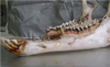
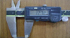
This study was an assessment of diagnostic accuracy. A total of 42 prefabricated titanium pins of similar length were inserted in seven dry sheep mandibles (Fig. 1). The length of these pins was measured before their insertion in the mandibles by using a digital caliper (Mitutoyo Corp., Kawasaki, Japan) with readability of 0.01mm(Fig. 2). To simulate soft tissue, mandibles were immersed in a water container and radiographed with NewTom VGi® (QR s.r.l., Verona, Italy). Imaging was carried out at a high-resolution field of view of 8 cm×12 cm with a voxel size of 0.125 mm and tube voltage of 120 kVp.
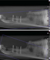
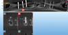
For the reconstruction of images by NNT viewer software (QR s.r.l., Verona, Italy), the orientation of slices was adjusted to parallel (i.e., 0°), +10°, +12°, -12°, and -10° with respect to the occlusal plane (Fig. 3). These intervals were chosen on the basis of a pilot study on the primary reconstruction of CBCT images conducted by the oral and maxillofacial radiology residents and faculty of our institution; each of these researchers had at least four years of experience with CBCT. This pilot study showed that the range of errors in angulation during image reconstruction was from -12° to +12°, and hence, five different angles within this range were chosen to evaluate the effect of the angulation error on the measurement outcomes. The images were viewed on a 20-inch Philips monitor (200P, Philips, Guildford, England) with a pixel resolution of 1024×1280 and color depth of 32 bits. Linear measurements of the lengths of pins were made by three oral and maxillofacial radiologists (Fig. 4). The observers were allowed to control the contrast, sharpness, and brightness of the images as well as the slice thickness and the interslice interval as desired. Each observer measured each length twice with a two-week interval between the measurements. The accuracy of linear measurements obtained by CBCT was reported using descriptive statistics and one-way analysis of variance (ANOVA); p<0.05 was considered statistically significant. Moreover, for the calculation of the inter-rater reliability accuracy in this study, a weighted kappa statistical analysis was used.
Results
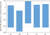
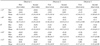
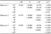
The difference in radiographic measurements made by the observers ranged from -0.64 to +0.06 at the orientation of -12°, -0.66 to -0.11 at -10°, -0.51 to +0.19 at 0°, -0.64 to +0.08 at +10°, and -0.64 to +0.1 at +12°(Fig. 5). Table 1 demonstrates the mean and standard deviation of the differences in measurements at each orientation considered and for each observer between the first and the second observations. The maximum and minimum mean absolute values of the error were 0.34 and 0.20 at the orientation of -12°, 0.37 and 0.27 at -10°, 0.18 and 0.14 at 0°, 0.31 and 0.23 at +10°, and 0.31 and 0.29 at +12°, respectively. The results showed that the mean absolute value of the error was greater at negative orientations than at the parallel position or at positive orientations of the slices with respect to the occlusal plane. The maximum mean absolute value of the error was 0.37±0.13 observed at the orientation of -10°, whereas the minimum mean absolute value of the error was 0.14±0.87 recorded at 0°. The observers underestimated the length of the pins at most of the orientations by 0.5 to 0.1mm(83.6%). In the second set of observations, taking into account the accuracy subgroups (error range), the reproducibility at all orientations was greater than 0.9. Table 2 shows the p value for the different orientation angles obtained using one-way ANOVA.
Discussion
The results of the present study revealed that by changing the slice orientation within the range of -12° to +12° with respect to the occlusal plane during image reconstruction, the accuracy of linear measurements using CBCT decreases; nevertheless, these measurements can still be considered highly accurate. In this study, titanium pins were used since titanium has a low atomic number and hence, will cause very small metal artifacts.9,10 Furthermore, the observers were allowed to change and adjust the contrast, sharpness, and gamma factor (which is among the features of the NNT viewer software) of the images, and by doing so, they could further minimize the metal artifacts.
The highest mean difference between measurements in this study was observed at the orientation of -10°, while the lowest mean difference was recorded at 0°, which implies that during image reconstruction, adjusting the slice orientation to be parallel to the occlusal plane results in reducing the error in the linear measurements.
The mean absolute error value at negative orientations was greater than at the parallel position or at positive orientations with respect to the occlusal plane. The maximum mean of the absolute value of the error was observed at the orientation of -10°, while the minimum mean absolute value of the error was recorded at 0°. However, the differences in the mean errors of measurements between the considered orientations (-12°, -10°, 0°, 10°, and 12°) and the gold standard was less than 1 mm. In 2011, Ganguly et al11 demonstrated that the acceptable measurement error for implant placement was less than 1 mm. Therefore, in our study, the accuracy of measurements at all orientations was considered clinically acceptable.
In a study by Sheikhi et al,12 the accuracy of linear measurements was compared at normal and at different head positions in a Galileos CBCT system. In their study, distances specified with radiopaque markers on the scans obtained at different head positions were measured. The mean difference between the physical (actual) and the radiographic measurements was found to be 0.45±0.05 mm.
Tomasi et al13 evaluated the reliability and the reproducibility of linear measurements in the mandible by using CBCT at two different orientations of 0° and 45°. The mean absolute error value was 0.4 mm, which is almost to the same as the values obtained in our study. Further, Fakhar and Abbaszadeh14 evaluated the effects of changing the mandibular plane orientation on the accuracy of the linear dimensions of the tomographic images. Analyses showed statistically significant differences between measurements made at the standard position and upon the upward tilt of the mandibular plane. In general, in most cases, the mean absolute error value was 1 mm or less, which is within the acceptable range. However, a downward tilt of the mandible caused a greater change in measurements than an upward tilt.
Hassan et al15 evaluated the accuracy of linear measurements performed using 3D CBCT images at two different head positions. The greatest difference in measurements observed between the 3D models and the gold standard was less than 0.5 mm, and no significant difference was found between the linear measurements at the different head positions. Moshiri et al16 compared the accuracy of linear measurements conducted using CBCT and conventional cephalograms and demonstrated that measurements using CBCT images had a higher accuracy than those using conventional lateral cephalograms. Further, they showed that the head position had no significant effects on the accuracy of linear measurements using CBCT.
Ludlow et al17 in their study on the NewTom 9000 machine revealed that the accuracy of linear measurements using CBCT was not significantly influenced by the variations in the head orientation while the image was being acquired.
Moshfeghi et al18 evaluated the linear measurement accuracy of CBCT using NewTom VG on 22 anatomic landmarks in four dry skulls. Fifteen linear measurements were obtained using a digital caliper. The skulls were scanned at two settings: (a) voxel size of 0.3 mm and (b) voxel size of 0.15 mm. Radiographic distance measurements were made using the NNT viewer software in the axial and coronal sections by three observers. A statistical analysis showed high inter- and intra-observer reliability.
In conclusion, changing the slice orientation in the range of -12° to +12° reduced the accuracy of linear measurements obtained using CBCT. However, the error values were smaller than 0.5 mm and were, therefore, clinically acceptable.
 XML Download