

 PDF
PDF ePub
ePub Citation
 Print
Print


Fibromyxoma is a synonym for odontogenic myxoma as reported in the World Health Organization (WHO) Classification 2005. It is a rare, benign, and locally aggressive odontogenic tumor of a mesenchymal origin,1,2 comprising 3-6% of all odontogenic tumors.3
Fibromyxoma of the jaw, either the mandible or maxilla, has been reported in the literature since the middle of the last century.4,5,6,7,8.9,10,11,12,13,14 Most cases occur in young patients mainly those in their 20 s or 30 s, and the condition has a female predilection. The mandible is more commonly affected than the maxilla, mainly in the posterior region. Usually, it causes a painless, slow-growing swelling.9,15
Radiographically, it has been demonstrated as a unilocular or a multilocular radiolucency in the tooth-bearing area with a well-defined border, but sometimes, it does not match this definition. Root resorption is rarely found in cases of fibromyxoma, and the scalloping margin between the roots occurs more frequently.3,6,12 Maxillary lesions may cause invagination of the maxillary sinus and expansion of its walls as revealed in cross-sectional imaging modalities such as computed tomography.13,14
Histologically, the tumor is composed of stellate and spindle-shaped cells with a haphazard arrangement in a loose mucinous or myxoid stroma, and its connective tissue may contain few collagen fibrils. Remnants of the odontogenic epithelium may also be present.13
This report describes a rare case of multiple fibromyxomas and highlights their clinical and radiographic features, including panoramic and magnetic resonance images, as well as histopathological features.
Case Report
A 13-year-old female patient visited the university hospital due to delayed eruption of some of her teeth. The cause of delayed eruption was not clear, but it might have been due to trauma, as when the patient was five, she had a severe car accident and her jaws were severely traumatized, but without any apparent jaw fractures. The deciduous teeth fell out, and the gingiva healed normally; however, there was no sign of any eruption of the permanent teeth afterwards. Surgery had previously been conducted to release the central incisors. No pain was reported by the patient except at the molar area while eating. There was no history of paresthesia. An unremarkable medical history was otherwise reported by the patient's guardian with normal previous laboratory investigations, namely, the complete blood count and the calcium, phosphorous, and alkaline phosphatase levels. In addition, the overall appearance of the patient was normal with no evidence of the presence of a systemic disease.
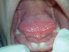
Clinical examination revealed multiple missing teeth with wide edentulous areas in both jaws except the anterior region and the first molar regions, and the covering mucosa was normal. Palpation showed slight obliteration of the buccal vestibule in the posterior region. The erupted teeth showed a normal color and architecture (Fig. 1). Neither facial asymmetry nor palpable lymph nodes were found on the extra-oral examination.
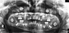
A panoramic radiograph revealed four pericoronal radiolucencies related to the four third molar regions. The four lesions demonstrated a well-defined border with a corticated margin, but this was more sharply and clearly detected in the mandibular lesions; the border of the lesions was not related to the cement-enamel junction of the related molars. The panoramic radiograph also revealed several impacted teeth with delayed development considering the patient's age (Fig. 2).
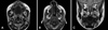
Magnetic resonance imaging (MRI) showed a homogenous intermediate signal intensity on the T1-weighted images and a heterogeneous slightly hyper-intense signal intensity on the T2-weighted images for the four lesions. Buccal and lingual cortical expansion could be detected with the mandibular lesions. Meanwhile, the maxillary lesions revealed invagination of the maxillary sinus (Fig. 3).
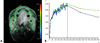
Dynamic MRI revealed a strong heterogeneous enhancement of the lesions. Time-intensity curves revealed rapid early enhancement followed by gradual enhancement and then, a slow and delayed washout (Fig. 4). Upon a diffusion MRI, the apparent diffusion coefficient (ADC) map revealed a low-to-intermediate heterogeneous signal, which indicates partially restricted diffusion.
Surgical removal of the four lesions was performed under general anesthesia. A histological examination revealed relatively hypocellular tumor tissues formed of spindle and stellate cells within a predominantly collagenous, focally myxoid background with intervening thin-walled vessels. The final diagnosis of the four lesions was fibromyxoma (Fig. 5).
Discussion
Although fibromyxoma is widely defined as a benign locally aggressive odontogenic tumor of the mesenchymal origin, but the nature of fibromyxoma is controversial. WHO Classification 2005 reported it as an equivalent term to odontogenic myxoma; at the same time, some authors have claimed that fibromyxoma is a subtype of odontogenic myxoma and comprises only a small percentage of it,13,16 while others have suggested that it was more related to odontogenic fibroma.17
The obvious issue is that fibromyxoma contains more fibers than odontogenic myxoma;18 however, the sequence of formation, whether it represents a myxomatous degeneration of odontogenic fibroma5 or collagen fiber production in odontogenic myxoma by fibroblast-like secretory cells,12,19 is unclear.20
Pericoronal radiolucent lesions are common in the mandible as well as the maxilla, and their differential diagnosis list includes dentigerous cyst, ameloblastoma, ameloblastic fibroma, keratocystic odontogenic tumor, adenomatoid odontogenic tumor, Pindborg tumor, and calcifying cystic odontogenic tumor. Odontogenic myxoma and odontogenic fibroma were recorded as rarities in pericoronal radiolucencies.21,22,23,24
Panoramic radiography was the way to discover the occult pericoronal lesions in the present case as well as the determination of their extension, shape and periphery, size, number, and effects on surrounding structures.25 Although the information acquired from the panoramic radiograph about the internal structure of the lesions that revealed a lack of calcifications and presence of impacted teeth inside was crucial, it could not provide evidence of the nature of the tissues composing these space-occupying lesions.
Aspiration aids in narrowing the differential diagnosis of pericoronal radiolucencies, but in some cases, aspiration may be impossible, particularly in inaccessible areas like the ramus of the mandible and the posterior region of the maxilla. Due to the superior contrast resolution of MRI, the detection of the solid or cystic nature of intra-bony lesions is possible. Unlike aspiration, MRI can reveal the solid or cystic composition in any part of the lesion and in the inaccessible areas.
MRI as a cross-sectional modality provides the chance to reveal the extension of the lesion in the orthogonal planes or any oblique planes as well as the effects on the surrounding structures. Furthermore, it can reveal the internal structure of the lesion; the intensity of the signal in the T1 and T2 spin echo images as well as the lack of peripheral enhancement in the dynamic MRI indicate that the lesion is not cystic in nature.
The intermediate and hyper-intense signals revealed by the T1 and T2 images, respectively, are similar to those of previous studies that performed MRI for odontogenic myxoma.26,27,28 In contrast to our findings, a study performed by Kawai et al in 199729 revealed an intermediate signal on the T2 images of odontogenic myxoma. This disagreement might be attributed to variations in the contents of the lesion, such as the ratio of fibrous to myxoid tissues, viscosity, protein concentration, hemorrhage, or hypocellularity.13
The results of dynamic MRI are in agreement with those of previous studies that used a contrast agent for the assessment of odontogenic myxoma cases regarding the pattern of enhancement.27,28 Our results are also quite similar to those of Asaumi et al in 200230 in that the current study and Asaumi et al showed a strong gradual enhancement followed by a slow washout, but at the same time, there are some differences between them where the current case showed rapid enhancement in the first few seconds before the gradual enhancement as well as the earlier washout demonstrated in our case. The heterogeneous appearances shown on the T2 images and the dynamic MRI indicate that the lesions were composed of more than one type of tissue, which was confirmed by the histopathological examination, and the partial restriction observed in the diffusion weighted images might refer to the fibrous components of the lesions.
To the best of our knowledge, no cases of multiple fibromyxomas of the jaws have been reported in the literature; only solitary cases have been reported. Multiple odontogenic fibromas are also extremely rare; only a few cases have been reported in the literature, and most of these were associated with enamel dysplasia and hypodontia.20,31,32
In conclusion, more cases and studies are needed to discover and document such unusual cases of multiple fibromyxomas related to the impacted teeth in the mandible and the maxilla as well as correlating this condition to the presence of multiple impacted teeth and delayed tooth development.
 XML Download