

 PDF
PDF ePub
ePub Citation
 Print
Print


Introduction
Diagnosis, treatment planning, and evaluation of treatment outcomes in endodontics are routinely done with the help of radiographs. Radiographic imaging is essentially two-dimensional (2D) imaging of a three-dimensional (3D) object. Furthermore, the interpretation of radiographs can be influenced by several confounding factors, including regional anatomy and superimposition of teeth and the surrounding dentoalveolar structures. The structures visualized by radiographs are also subject to geometric distortions.1 These problems can be overcome by the use of 3D imaging using computed tomography (CT).
The application of CT scans in endodontics was first reported by Tachibana and Matsumoto in 1990.2 A CT scan uses a fan-shaped beam and multiple exposures around an object to reveal the internal architecture of this object, thereby helping the clinician to view morphologic features as well as pathology from different 3D perspectives. The distinct advantage of a CT scan is that it allows for a 3D reconstruction of root canal systems. CT scanning has been suggested as the preferential imaging modality in difficult situations demanding the localization and description of root canal systems because of its ability to render 3D information.3 The dental CT, also called DentaScan (GE Healthcare), was first reported by Schwartz et al.4 The objective of this article is to review the applications of different modalities of CT in endodontics. CT scans play an important role in the analysis of root canal anatomy, detection of apical periodontitis, diagnosis of trauma, and presurgical assessment.
Three types of CT scans, namely cone-beam CT (CBCT), spiral CT, and peripheral quantitative CT (pQCT), have been used in endodontic research to study the root canal anatomy and its variations.
CBCT scanning or digital volume tomography (DVT) uses an extraoral imaging scanner to produce 3D scans of the maxillofacial skeleton at a considerably lower radiation dose than that required for conventional CT scanning. CBCT scanning has been shown to be more accurate than digital radiographs in identifying root canal systems. CBCT scanning has also been used in vivo in diagnosis and preoperative assessment for root canal treatment.5 CBCT differs from medical CT imaging, in that the entire set of 3D volume data is acquired in the course of a single sweep of the scanner, using a simple, direct relationship between the sensor and the source. The X-ray beam is cone-shaped and captures a cylindrical or spherical volume of data, described as the field of view.
Another CT technique, pQCT, was originally introduced for bone mineral analysis. The unit works with a specially developed X-ray tube having a minute focal spot, whilst the detector system consists of a series of miniature semiconductor crystals. The device is equipped with a special detector collimator that can be switched among up to four collimator sizes corresponding to the four section thicknesses (100, 250, 500, and 750 µm). Although the planar resolution of pQCT (70 µm×70 µm) does not have the same resolution as micro-CT, it might provide a nondestructive morphological investigation at a low cost and relatively short scanning time. A report on the use of pQCT in the study of root canal anatomy showed that this method offers an accurate 3D reconstruction of the root canal systems and analysis of endodontic procedures.6
Spiral CT has been used in several cases of the diagnosis of aberrant root canal systems,7,8 as well as in the identification of the root canal morphology of Indian molars.9 Spiral CT or volume acquisition CT employs simultaneous patient translation through the X-ray source with a continuous rotation of the source-detector assembly. The raw projection data are acquired with a spiral sampling locus in a relatively short period. These data can be viewed as conventional transaxial images such as multiplanar reconstructions or as 3D reconstructions.
Analysis of root canal anatomy
Successful endodontic therapy stems from thorough canal debridement and effective filling of the root canal system, for which knowledge of the morphology of root canals is a critical prerequisite.10 The knowledge of root canal systems gained from in vitro studies helps in understanding the racial predisposition of different canal anatomies.11,12 The methods most commonly used for analyzing the root canal morphology are canal staining and tooth clearing,13 conventional radiographs,14 digital and contrast medium-enhanced radiographic techniques,15 radiographic assessment enhanced with contrast media,16 and more recently, CT techniques.17,18 An ideal technique would be one that is accurate, simple, non-destructive, and, most importantly, feasible in an in vivo scenario.
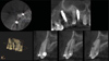
A recent study compared the efficacy of three types of CT scans (namely, CBCT, spiral CT, and pQCT) and digital radiographs (plain and contrast medium-enhanced) in identifying root canal systems in vitro. The canal staining and clearing technique was considered the gold standard. The evaluators failed to identify one or more root canals with digital radiographs in 23.8% of the teeth, contrast medium-enhanced digital radiographs in 14.8%, spiral CT in 15.58%, CBCT in 0.29%, and pQCT in 2.05% of the teeth. It was concluded that CBCT was the most accurate CT technique for identifying root canal systems in endodontics.19 The difference between these methods with respect to the identification of the root canal systems is mainly because of the variations in the slice thickness. CBCT has a slice thickness of 80-200 µm, while pQCT and spiral CT have slice thicknesses of 250 µm and 650-1000 µm, respectively.(Fig 1)
Several studies have reported root canal anatomical variations as a function of racial divergences. Liu et al studied the canal morphology of Chinese mandibular central incisors using CBCT and noted that in the population studied, the incidence of two canals in the lower central incisor was very low.20 Similarly, the anatomical variations in maxillary and mandibular teeth have been studied in different races, and with the application of CT for this purpose, accurate data are available for clinical use.20,21,22,23,24
Oshishi et al25 demonstrated the root anatomy and canal morphology of paramolar tubercles using CT. This was the first report to demonstrate the clinical relevance of CT in diagnosing the importance of the paramolar tubercle in endodontics. It was shown that the root canal of the tubercle united with the distobuccal root. An analysis of root canal geometry by CT was also reported by Peters et al.26 A micro-CT with a cubic resolution of 34 microns was used to assess the root canals of maxillary molars. Their study reported specific variables and indices that could be used for further studies on root canal anatomy. CT was also shown to be comparable to histology (the gold standard) in detecting root canal anatomy and morphology in the mesiobuccal roots of the maxillary first molars.27
In a study comparing tuned aperture computed tomography (TACT) with conventional D-speed films to identify root canals in the extracted human molars, Nance et al28 reported that TACT detected a fourth canal in the maxillary molar and a third canal in the mandibular molars, 36% and 80% of the time, respectively. In contrast, conventional films did not detect these extra canals in any of the samples. Another study showed contradictory results. Barton et al29 compared the parallax with pairs of conventional directexposure film intraoral radiographs (both D-speed and F-speed), parallax with pairs of charge-coupled device-based digital images acquired through the use of Trophy RVG UI, and charge-coupled device-based images acquired through the use of a Trophy RVG UI sensor and tomosynthetically reconstructed by the TACT Workbench software. The study showed that there was less than a 40% chance of locating the second mesiobuccal canal (MB2) in the mesiobuccal root of the maxillary first molar teeth by using parallax with pairs of digital or analog radiographs. Furthermore, TACT did not significantly affect the rate of detection of MB2 canals (37.9%) as compared to conventional films (39.2%) and RVG UI (39.6%).
The influence of age on the size and shape of the pulp cavity of premolars was assessed using micro-CT. The researchers employed 3D reconstruction software to show that the pulp cavity size decreased with an increase in age.30
Assessment of root canal preparations
Traditionally, hand instruments were used to shape root canals. Contemporary endodontics has available several automated techniques, of which those involving the use of rotary and reciprocating nickel-titanium instruments are the most commonly used in clinical practice. Prior to the clinical introduction of these instruments and to understand the clinical impact of these instruments on root canal shaping, the geometry of root canals prepared by these nickel-titanium instruments has to be evaluated. Peters et al31 were the first to employ a CT-based technique (micro-CT) to analyze the efficacy of different techniques of root canal preparation. They concluded that micro-CT was an indispensable tool in the research on the effectiveness of preparation instruments and techniques in vitro. Numerous studies have been performed since then, using various types of CT to determine the efficacy of root canal instruments. Although most studies employed micro-CT, few works have employed CBCT as well. Considering the differences in the slice thickness and the resolution of micro-CT and CBCT, the results of studies employing CBCT for this parameter might be questionable. Sonntag et al32 reviewed the evaluation of root canal curvatures before and after root canal preparation. It was realized that only graphical representation and mathematical formulae existed for the analysis of root curvatures. However, these might not be deemed accurate. It was reported that CT-based 3D reconstructions could offer an extensive assessment of root canal preparation in a non-destructive manner.33
The introduction of CT has allowed the detection of C-shaped canals in several studies, thereby providing a valuable database to endodontic practice and radiology. In a sample size of 491 mandibular molars, C-shaped canals were located in 8.1% using spiral CT. Using 3D reconstructions along with spiral CT, the authors were also able to classify the morphology of C-shaped canals by using Manning's criteria.34
Detection of periradicular lesions
CT scans, and more importantly CBCT, can detect radiolucencies of the cortical bone even when they cannot be visualized on conventional radiographs. CBCT can also detect bone defects of the cortical bone and cancellous bone separately, in contrast to radiographs wherein lesions must erode the cortex before they can be visualized. Estrela et al35 demonstrated that the prevalence of apical periodontitis was significantly higher when CBCT was used as the diagnostic method. Their study showed that CBCT scans detected periapical lesions in 62% more cases than radiographs. Furthermore, it has also been shown, with histological controls as the gold standard, that CBCT is more sensitive in identifying apical periodontitis.36
Earlier detection of periradicular radiolucent changes with CBCT should result in an earlier identification and management of endodontic disease; this in turn should result in a better outcome from endodontic treatment as teeth can be treated sooner. In situations where patients have poorly localized symptoms associated with an untreated or previously root-filled tooth and clinical and periapical radiographic examinations show no evidence of disease, CBCT might be indicated to detect the presence of previously undiagnosed periapical disease. CBCT has been found to have 100% sensitivity (1.0) and specificity (1.0) in the detection of artificially created periapical lesions in dry human mandibles.36
Diagnostic dilemmas are also common in periapical radiolucencies. Benign and malignant lesions like carcinoma, odontogenic cyst, and periapical cemental dysplasia might mimic periapical lesions on a radiograph. Advanced imaging techniques are more useful than radiographs in the diagnosis of these lesions. Sekerci et al37 reported two cases where CBCT was employed to diagnose an anatomical variation of the maxillary sinus that mimicked a periapical cyst, thereby avoiding unnecessary intervention.
Presurgical assessment
Three-dimensional imaging allows clear identification of the anatomical relationship of the root apices with important neighboring anatomical structures such as the inferior dental canal, mental foramen, and maxillary sinus. CBCT may play an important role in the periapical microsurgery of palatal roots of the maxillary first molars. The distance between the cortical plate and the palatal root apex can be measured, and the presence or absence of the maxillary sinus between the roots can be assessed.38
The following parameters can be easily assessed using CT scans before microsurgical treatment planning: proximity to critical vital structures, thickness and architecture of the cortical plate and the cancellous bone, inclination of roots, root canal morphology, untreated root canals, cracks and fractures, and the location and extent of periapical pathoses. The likelihood of detecting periapical lesions with periapical radiographs has been shown to be reduced when the root apices were in close proximity to the floor of the maxillary sinus and when there was <1 mm of bone between the periapical lesion and the sinus floor. Furthermore, confident access to the surgical site can be planned by rapid prototyping and stereolithiography.39
The diagnosis of root resorptions is a challenging issue in endodontics. Radiographs often fail to detect root resorptions along the lingual aspects of teeth. Furthermore, the cervical burnout, which is characteristic of periapical radiographs serves as a confounding factor in diagnosing resorptive defects. CBCT-reconstructed images have been successfully used in the diagnosis and management of resorptive lesions.40 One can detect the nature and location of the resorptive defect, in addition to identifying the portal of entry of these lesions. Treatment strategies can be easily planned on the basis of the 3D imaging of these defects.41
Diagnosis of dental trauma
A single scan followed by multiplanar views can be used to assess the severity of dental trauma. This is particularly useful in luxation injuries and in fractures of the dentoalveolar complex. Root fractures in the horizontal direction need multiple angulated radiographs to detect the nature of fracture. Despite this, they may not be completely identified. With CT scans (preferably CBCT), horizontal root fractures can be identified with ease.42 CBCT also has an added advantage in trauma cases in that it is an extraoral technique and hence, more comfortable for the patient. CBCT scans are also able to detect cortical bone fractures, which cannot be diagnosed from a clinical or conventional radiographic examination.
Vertical root fractures (VRF) offer several diagnostic challenges. The lack of consistent signs and symptoms as well as the low sensitivity of conventional radiographs make the detection of VRF difficult. CBCT has been shown to be an important tool in the diagnosis of VRF.43 Edlund et al44 determined the diagnostic accuracy of the CBCT detection of suspected VRFs in endodontically treated teeth by using exploratory surgery to confirm the presence or absence of a fracture. Their study revealed that the positive predictive value and the negative predictive value of CBCT was 91% and 67%, respectively. With a sensitivity of 88% and specificity of 75%, CBCT proved to be a superior diagnostic tool in the detection of VRF.
Metska et al45 evaluated two limited field-of-view scanners (NewTom 3G and 3D Accuitomo 170) and found that the sensitivity, specificity, and accuracy of the NewTom 3G were 75%, 56%, and 68%, respectively, and for the 3D Accuitomo 170, they were 100%, 80%, and 93%, respectively. The positive predictive value and the negative predictive value were 75% and 55%, respectively, for NewTom 3G and 90% and 100%, respectively, for 3D Accuitomo 170. The results of their study were interesting with respect to clinical practice because the reproducibility and accuracy of VRF detection appeared to depend on the CBCT system used. Also, the sensitivity of the NewTom 3G system was reduced by the presence of metallic posts in contrast to the i-CAT system (Imaging Sciences International, Hatfield, PA, USA).45
The favorable voxel size of CBCT (0.125-2 mm) and the ability to achieve multiplanar image reconstruction in the axial, sagittal, and coronal sections, in addition to the minimal artifact interference make this modality extremely important in the assessment of maxillofacial structures. However, root-filling materials might serve as artifacts.46 This in addition to beam hardening during CBCT might result in an erroneous diagnosis of VRF. Bechara et al47 demonstrated that CBCT with smaller fields of view had higher accuracy and sensitivity for detecting VFR than CBCT with larger fields of view as well as photostimulated phosphor plates.
Discussion
Treatment success in endodontics (non-surgical and surgical) results from an accurate diagnosis and debridement of the root canal space. High-end imaging techniques must be employed in endodontics for making a correct diagnosis and to provide the best standard of care. Three types of CT scans, namely CBCT, spiral CT, and pQCT, have been used in clinical endodontics. In vitro research in endodontics commonly employs micro-CT.
The application of 3D imaging methods in clinical endodontics should be based on a benefit-risk analysis. The importance of CT scans, particularly CBCT, in the diagnosis of root canal anatomy, root resorption, root fractures, and differential diagnosis of periapical lesions has been reviewed in this article. The advantages of CT scans with respect to these factors are clear. Nevertheless, it must be understood that CT scans use ionizing radiation and patient exposure should be kept as low as reasonably practicable, based on the As Low As Reasonably Achievable (ALARA) principle. Longer scanning times and high costs may prohibit the routine use of CT scans in endodontics. However, considering the potential advantages of CT scans in endodontics, more economical systems should be developed so that 3D imaging becomes an integral part of endodontic diagnostics.
 XML Download