Abstract
A ranula is a bluish, transparent, and thin-walled swelling in the floor of the mouth. They originate from the extravasation and subsequent accumulation of saliva from the sublingual gland. Ranulas are usually limited to the sublingual space but they sometimes extend to the submandibular space and parapharyngeal space, which is defined as a plunging ranula. A 21-year-old woman presented with a complaint of a large swelling in the left submandibular region. On contrast-enhanced CT images, it dissected across the midline, and extended to the parapharyngeal space posteriorly and to the submandibular space inferiorly. Several septa and a fluid-fluid level within the lesion were also demonstrated. We diagnosed this lesion as a ranula rather than cystic hygroma due to the location of its center and its sublingual tail sign. As plunging ranula and cystic hygroma are managed with different surgical approaches, it is important to differentiate them radiologically.
Ranulas originate from the extravasation and subsequent accumulation of saliva from the sublingual gland. If a salivary duct is obstructed, secretory back-pressure builds leading to a duct rupture with mucus being forced into the surrounding tissues. The origin of the ranula was unknown until toward the end of the twentieth century, when some authors concluded that the ranula arose from the sublingual gland.1,2 The sublingual gland is a spontaneous secretor and produces a continuous flow of mucus even in the absence of nervous stimulation.3 Ranulas typically have a bluish appearance and a fairly well-circumscribed, soft, painless, fluid-containing intraoral swelling. Most of the patients with ranula present with a gradually enlarging swelling of the floor of the mouth. The swelling is round or oval, and fluctuant. An intraoral swelling accompanied by a submandibular, cervical, and parapharyngeal extension is often defined as plunging ranula.4 CT scanning plays an important role in the diagnosis of a ranula.5-7 While most simple ranulas involve the sublingual space, the plunging ranula extends to the parapharyngeal space and the cervical space. In rare cases, a plunging ranula can have a subtle septation, which is usually related to a previous surgical treatment or traumatic history. The present report described a rare case of a giant plunging ranula with several septa and fluid-fluid levels.
A 21-year-old woman visited our department complaining of a large painless swelling in the left submandibular region. The swelling had been recognized at its sudden onset two months earlier. Intraorally, her mouth floor was bluish and elevated. On palpation, the swelling revealed a soft, painless, and fluid-containing mass. The patient had no traumatic or surgical history, and the swelling did not cause difficulty in swallowing or speaking. Routine blood tests and the thyroid profile were within normal limits.
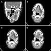
Panoramic radiograph revealed no pathological changes. Contrast-enhanced computed tomography (CT) scan demonstrated a large rim-enhanced fluid attenuation mass occupying both sublingual spaces with an anterior connection (Fig. 1). The lesion extended into the left parapharyngeal space superiorly and compressed the left submandibular gland inferiorly (Fig. 1A). Anteriorly, it extended to the right sublingual space in a horseshoe shape (Fig. 1B). At the lower level of the lesion, several linear septa were noted (Fig. 1C). A fluid-fluid level, which is the interaction between two fluids with different viscosities, was also noted (Fig. 1D). Although the septation and fluid-fluid level within the lesion made the differential diagnosis from a cystic hygroma difficult, considering the location of the lesion in the sublingual space, it was diagnosed as a plunging ranula.
Under general anesthesia, an incision was made in the left lingual vestibule, and excision of the lesion along with extirpation of the left sublingual gland was performed. At surgery, the cystic lesion was found to be filled with a viscous and yellowish mucous fluid. After removal of the left sublingual gland, a cut-down tube was inserted into the middle portion of the left Wharton's duct.
The histopathologic examination of the specimen from the sublingual gland revealed ruptured acinar cells (Fig. 2). The patient made an uneventful recovery. The cut-down tube inserted into the left Wharton's duct was removed after 2 weeks. The patient has not experienced a recurrence 6 months postoperatively.
The descriptive term ranula normally refers to a bluish, transparent, thin-walled swelling in the floor of the mouth. While simple ranulas are all confined to the sublingual space, plunging ranulas are centered on the submandibular space and tend to spill into one or more adjacent spaces.7 The nature of ranulas was unknown until 1956, when Bhaskar et al8 investigated the pathogenesis histopathologically and experimentally, and they concluded that ranula was produced by the extravasation of saliva from a damaged salivary sublingual gland and was not lined by epithelium. The occurrence of ranula is rare, and the reported male-to-female ratio is 1 : 1.3, without significant side preference.9 The plunging ranula most frequently occurs in the second and third decades of life, with an age range from 3 to 61 years, and most commonly involves the right side.10,11
Most giant plunging ranulas are located from the ipsilateral sublingual space to the parapharyngeal space. However, the present case of plunging ranula passed the midline and extended to the contralateral sublingual space. Based on our review of the English literature, only two cases of huge plunging ranula which passed the midline have been reported.11,12 The previous cases which passed the midline showed an extension into the cervical space inferiorly, but the present case did not extend beyond the submandibular gland. A rare case of plunging ranula with subtle septa was discussed. These septa might have been secondary to surgical procedures or a traumatic history. In those cases, wall thickening and minimal internal septa formation were noted.13,14 A trauma or a surgery might lead to the development of scar tissue, and it might be detected as a subtle septation within the plunging ranula. In the present case, contrast enhanced CT images demonstrated several mild and subtle septa. Although no previous trauma or surgery was found by history taking, it might be expected that the patient was unaware of the previous trauma considering the fluid-fluid level within the lesion. A fluid-fluid level has, in rare cases, been reported to occur in the plunging ranula and refers to the interaction between two fluids with different viscosities, commonly by hemorrhage.13 The blood vessels near the submandibular gland area are weak; therefore, the fluid-fluid level might easily result from trauma to the area.
The differential diagnosis for the present case included a cystic hygroma. Cystic hygroma rarely presents in the sublingual space, but if present, it reveals very similar images on CT scan to those of a plunging ranula. For cystic hygromas, the mainstay of treatment is surgical excision. The surgical team should attempt to completely remove the lymphangioma or to remove as much as possible, sparing all vital neurovascular structures.15 Treatment of a plunging ranula is also surgical excision, but the difference is the complete excision of the sublingual gland. There are several treatment options for ranula, which include excision of only the ranula and marsupialization. However, excision of the entire sublingual gland and ranula reported the lowest recurrence rate of 1.55%. As plunging ranula and cystic hygroma are managed with different surgical approaches, the differential diagnosis is critical.14 Cystic hygroma arises from the abnormal development of fetal lymphatic tissue. It typically presents early in life, with 50% present at birth and 90% evident by 2 years of age. Cystic hygroma is characteristically infiltrative in nature and typically involves contiguous anatomic regions in the neck. It appears as defined but poorly circumscribed, lobulated, septated, homogeneous, and with fluid attenuation on CT images. A previously infected lesion may show heterogeneous attenuation due to previous hemorrhage or high protein content. In case of hemorrhage, fluid-fluid levels may be observed, as with the present case. Therefore, the differential diagnosis between plunging ranula and cystic hygroma is often difficult on imaging. The characteristics of cystic hygroma for lobulation, strong septation, and rarity in the sublingual space should be helpful for diagnosis. In our case, the histopathologic examination demonstrated ruptured acinar cells of the sublingual gland, which confirmed the diagnosis of the plunging ranula.
For a ranula, surgery is the first-choice of treatment. The recurrence rates of ranula were not related to the swelling patterns and surgical approaches, but intimately related to the methods of surgical procedure.16 Effective treatment is removal of the involved sublingual gland or inducing sufficient fibrosis to seal the leak through which mucus extravasates.17 In the present case, the plunging ranula and ruptured sublingual gland were removed and the patient showed no recurrence 6 months postoperatively.
In conclusion, as plunging ranula and cystic hygroma are managed with different surgical approaches, it would be important to differentiate them radiologically. Although a submandibular cystic lesion with septa and a fluid-fluid level would be unusual for a ranula, a plunging ranula should be considered in the differential diagnosis along with a cystic hygroma.
Figures and Tables
 | Fig. 1Contrast-enhanced CT images show a large insinuating, rimenhanced fluid collection occupying both sublingual spaces. A. A coronal contrast-enhanced CT image shows the superior extension of the lesion into the parapharyngeal space and inferior displacement of the left submandibular gland by the lesion. B. An axial contrast-enhanced CT image demonstrates a horseshoe shaped fluid attenuation lesion. C. A contiguous axial contrast-enhanced CT image at a level slightly inferior to (B). Note several subtle septa within the lesion. D. A contiguous axial contrast-enhanced CT image at a level slightly inferior to (C) reveals a fluid-fluid level. |
References
1. Catone GA, Merrill RG, Henny FA. Sublingual gland mucusescape phenomenon-treatment by excision of sublingual gland. J Oral Surg. 1969. 27:774–786.
3. Harrison JD, Fouad HM, Garrett JR. Variation in the response to ductal obstruction of feline submandibular and sublingual salivary glands and the importance of the innervation. J Oral Pathol Med. 2001. 30:29–34.

4. van den Akker HP, Bays RA, Becker AE. Plunging or cervical ranula. Review of the literature and report of 4 cases. J Maxillofac Surg. 1978. 6:286–293.
5. Koeller KK, Alamo L, Adair CF, Smirniotopoulos JG. Congenital cystic masses of the neck: radiologic-pathologic correlation. Radiographics. 1999. 19:121–146.
6. Rho MH, Kim DW, Kwon JS, Lee SW, Sung YS, Song YK, et al. OK-432 sclerotherapy of plunging ranula in 21 patients: it can be a substitute for surgery. AJNR Am J Neuroradiol. 2006. 27:1090–1095.
7. Kurabayashi T, Ida M, Yasumoto M, Ohbayashi N, Yoshino N, Tetsumura A, et al. MRI of ranulas. Neuroradiology. 2000. 42:917–922.

9. Chidzonga MM, Mahomva L. Ranula: experience with 83 cases in Zimbabwe. J Oral Maxillofac Surg. 2007. 65:79–82.

12. Davison MJ, Morton RP, McIvor NP. Plunging ranula: clinical observations. Head Neck. 1998. 20:63–68.

13. Coit W, Harnsberger H, Osborn A, Smoker W, Stevens M, Lufkin R. Ranulas and their mimics: CT evaluation. Radiology. 1987. 163:211–216.

14. Macdonald AJ, Salzman KL, Harnsberger HR. Giant ranula of the neck: differentiation from cystic hygroma. AJNR Am J Neuroradiol. 2003. 24:757–761.
15. Burezq H, Williams B, Chitte SA. Management of cystic hygromas: 30 year experience. J Craniofac Surg. 2006. 17:815–818.


 PDF
PDF ePub
ePub Citation
 Print
Print


 XML Download