

 PDF
PDF ePub
ePub Citation
 Print
Print


Multiple calcifying hyperplastic dental follicle (MCHDF) is an extremely rare condition characterized by multiple impacted teeth and enlarged dental follicles that contain abundant calcifications and rests of odontogenic epithelium. A PubMed search with the keywords "calcification AND dental follicle" and "hyperplastic dental follicle" made in May 2013 and examination of the references of the relevant articles revealed that, up to that date, 11 cases had been reported.1-6 Two of these cases were originally published as odontogenic fibroma-like cases.2,4 Later, when Gardner and Radden1 analyzed these cases together with two additional cases, they suggested the term MCHDF and proposed that the condition is sufficiently distinctive to be considered a pathologic entity.
In all of the reported cases, the patients were male and younger than 40 years. The impacted teeth were mostly third or second molar, canine, or second premolar teeth, and they were associated with pericoronal radiolucencies delineated by sclerotic borders.1-6 In three cases, focal discrete radiopacities within the pericoronal radiolucencies were detected on the radiographs; however, calcification in the enlarged dental follicles was a significant histopathologic feature of this entity.1-3
The purpose of the present report was to describe the clinical, radiographic, and histopathologic features of a case of MCHDF.
Case Report
A 31-year-old female visited our clinic with the complaints of pain and gingival swelling on the left maxillary alveolar region and consequent difficulty of eating. Her dental history revealed that she had a dental visit due to her carious and crowded teeth when she was 17 years old. Her carious molar teeth had been extracted and she had applied for orthodontic treatment. The palatally positioned left maxillary incisor teeth were treated for a four-year period. At that time, her impacted teeth were diagnosed on routine radiographs, and she had a surgical operation for the extraction of one of her impacted right mandibular teeth at the age of 18. Her orthodontist followed up on the eruption of the other impacted teeth yearly until she was 21 years old, but then she interrupted her dental treatment and follow-up visits until our examination.
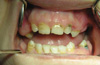
The patient had also been diagnosed with hypothyroidism when she was five years old. She was undergoing levothyroxine sodium (Levotiron®, Abdi İbrahim, Istanbul, Turkey) therapy, and her T3, T4, and TSH hormone levels were normal. On extraoral examination, she was approximately 1.50 meters tall and had sparse hair. Intraoral examination revealed missing teeth, diastemata, macroglossia, and buccolingual expansion of the alveolar bone (Fig. 1).
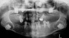
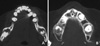
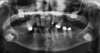
A panoramic radiograph revealed six impacted permanent teeth: the third molar teeth in all quadrants and the maxillary canine teeth. The crowns of these impacted teeth were surrounded with cyst-like lesions containing radiopaque foci, and well-defined corticated borders. The impacted mandibular molar teeth were located in the apical direction near the inferior cortex of the mandible, and the impacted maxillary teeth were located close to the floor of the maxillary sinus (Fig. 2). For further information, computed tomography was performed. Axial sections of the mandible revealed rounded radiolucent lesions associated with the crowns of the right and left mandibular molar teeth. These lesions contained radiopaque foci and were interpreted as calcification (Fig. 3A). In axial sections of the maxilla, the mixed appearance of the lesions with well-defined borders was noteworthy (Fig. 3B).
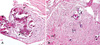
After surgically exposing the impacted left mandibular third molar tooth, an incisional biopsy specimen was obtained from the center of the lesion, under direct vision. Subsequent to biopsy, histopathological examination of the specimen obtained from the follicle of the tooth was made. Microscopic examination revealed loose to moderately dense collagenous connective tissue with abundant calcified material and sparse epithelial islands. The calcifications were mostly acellular type 1 calcification but scanty cellular type II calcifications were also detected. Inflammatory cell infiltration was not observed (Fig. 4). A diagnosis of dental follicles with calcifications was made.
The patient refused a surgical operation for removal of the impacted teeth, and she was scheduled for a follow-up visit. One year later, there was not any alteration in the radiographic appearance of the lesions, and then the patient was lost to follow-up. Five years later, the patient showed up for a follow-up appointment. A new panoramic radiograph revealed slight enlargement of the calcified follicles and an increase in the density of the calcifications. A focal increase in the radiopacity associated with the crown of the right mandibular third molar tooth, a small radiopaque prominence associated with the inferomedial aspect of the follicle of the left mandibular third molar tooth, and irregularity of the contours of the hyperplastic follicles were detectable on the radiograph made five years after initial presentation (Fig. 5). The patient refused any surgical operation or prosthetic restoration again.
Discussion
MCHDFs are atypical follicles or hamartomatous anomalies.2 However, the lesions radiographically and histologically resemble many other benign neoplasms and may pose diagnostic problems for clinicians including maxillofacial radiologists and pathologists who are not familiar with this condition.2,4,6,7
The crowns of unerupted teeth are normally surrounded by a dental follicle, and radiographically, the dental follicle appears as a radiolucent halo that has a thin radiopaque border. The follicular spaces of long-standing impacted teeth are frequently diminished, with the exception of maxillary canine teeth having enlarged follicles.8 Histopathologic examination of an enlarged dental follicle sometimes reveals no pathosis, while at the same time, radiographically normal dental follicles may exhibit histopathological changes.9,10 Despite the controversy in the literature regarding the establishment of criteria for the differentiation of incipient pathoses and enlarged dental follicles, the width of the pericoronal space has been used as a criterion to distinguish between a normal and an abnormal follicle. When the follicular radiolucency reaches 2.5 mm in width on the periapical and 3 mm on the panoramic radiograph, pathosis is suggested, and if the radiopaque border, which is the radiographic image of the surrounding cortical plate, is not well-defined, this is also a sign of pathologic change.8 In the present case, all of the follicular spaces of the impacted teeth exceeded 3 mm in diameter, and the cortical plates around the right mandibular and maxillary third molar and the left maxillary canine teeth were well defined, whereas the right maxillary canine, left maxillary molar, and mandibular third molar teeth were surrounded by roughly well-defined radiopaque borders, suggesting pathosis.
Radiographically, calcifying cystic odontogenic tumor, adenomatoid odontogenic tumor (AOT), ameloblastic fibroodontoma, and calcifying epithelial odontogenic tumor (CEOT) may show radiopaque foci, and they may be associated with unerupted teeth.7,11-13 Another rare entity that shows radiopaque foci and may be associated with unerupted teeth is MCHDF.7
Calcifying cystic odontogenic tumor tends to occur anterior to the first molar and is especially associated with cuspids and incisors, and most AOTs occur in the anterior maxilla, especially the canine region.14 In our case, only two of the enlarged follicular spaces containing radiopaque foci were associated with canine teeth whereas there were four others associated with molar teeth, and slow-growing, painless jaw swelling was absent, which was reported to be a feature of these two pathologic entities. Ameloblastic fibro-odontoma and CEOT both have a predilection for the posterior mandible; however, ameloblastic fibro-odontoma occurs predominantly in children and young adults, and jaw expansion in CEOT is a regular feature but was not observed in the present case.7,12,15
Calcifying cystic odontogenic tumor, AOT, ameloblastic fibro-odontoma, and CEOT are mostly encountered as solitary lesions, and multifocal cases are very rarely reported.16,17 Therefore, when multiple impacted teeth show enlarged follicular spaces containing radiopaque foci, MCHDF should be considered.1-3
Including the case presented, the average age of all reported cases is 20.1 with a range of 11-40 years. A review of the previous cases revealed no other report of this phenomenon in females.1-6 Clinically, diastemata and buccolingual expansion of the alveolar bone, which were also features of our case, were observed in one previously reported case, and in another report, firm, well localized asymptomatic expansions were noted in the mandibular and maxillary premolar regions.2,5 The vast majority of the involved teeth were the canines, second and third molars, and premolars.1-6 In the present case, the impacted teeth were also the third molar and canine teeth. Congenitally missing or supernumerary teeth were previously reported but, at present, no other dental anomaly was known to be associated with MHCDF.4,5 In the present case, there were missing teeth and a history of extraction of carious molar teeth, but it was not definitely known if there were any congenitally missing teeth or extracted supernumerary teeth.
Recently, Cho et al5 reported five MCHDF cases; all of the patients had initially visited local clinics for the evaluation of delayed eruption of permanent molar teeth. According to the initial evaluation, all of the panoramic radiographs showed multiple odontogenic cysts, and the radiologists originally interpreted the enlarged follicles as dentigerous cysts.5 Also, in other cases, the radiographic appearances of the lesions were described to be radiolucent areas around the crowns of the unerupted teeth.1-4,6 The authors did not mention any radiopacities associated with the enlarged dental follicles. On the other hand, in three of the reported cases and in the case presented here, focal discrete radiopacities were detected within the pericoronal radiolucencies.1-3 Dare et al18 stated that intraoral periapical radiographs enabled the detection of the radiopacities in adenomatoid odontogenic tumor within radiolucency even with minimal calcified deposits, and panoramic radiography was often unable to demonstrate radiopacities in adenomatoid odontogenic tumor when the calcification was minimal. Therefore, when radiolucent lesions are encountered on panoramic radiographs, periapical radiography should be useful to detect radiopaque foci, if present. Follow-up radiographs taken in one patient revealed a slight increase in calcification within some of the pericoronal lesions.2 The differences in exposure parameters and patient positioning make it difficult to compare the panoramic radiographs; however, radiographic examination in the present case also revealed that the calcification process and follicular hyperplasia were continuing. The alterations were not detectable on the first follow-up radiograph made one year after diagnosis but became prominent five years later. Therefore, long-term follow-up is suggested for untreated cases.
In our case, microscopic examination revealed various spherical calcifications in the fibrous connective tissue and the histopathologic diagnosis was dental follicle containing calcifications. The histopathological features were abundant calcifications and rests of odontogenic epithelium in an enlarged dental follicle. It was not unusual to find inactive rests of odontogenic epithelium and calcified droplets of cementum-like material in follicles of normally developing or unerupted teeth, which have been associated with the induction of the dental ectomesenchyme.19 The distinct feature of our case was multiplicity of unerupted teeth. The radiographic features were suggestive of MCHDF, and the histopathological features, hyperplastic dense fibrous connective tissue with numerous deposits of type I and type II calcifications and rests of odontogenic epithelium, observed in the present case were identical to the previous cases of MCHDF.1,2,5
The mesenchymal cells of the dental follicles play an important role in the signalling pathway to induce eruption.20 Defective regulation of matrix metalloproteinases mediating connective tissue remodelling in non-syndromic hyperplastic dental follicles was suggested as a cause of abnormal tooth eruption.21 Thus, retarded eruption or impaction of teeth may be associated with the calcifying phenomenon that originates from the mesenchymal cells. According to Gardner and Radden,1 there was no apparent relationship of the present condition to either cleidocranial dysplasia22 or to Gardner's syndrome,23 both of which involve multiple impacted teeth. Hypothyroidism (juvenile), which was also a feature of our case, is a condition related to delayed eruption.14,24 In our case, it could be speculated that hyperplastic follicles hindered eruption of the involved teeth, which were probably delayed in eruption because of hypothyroidism. On the other hand, hypothyroidism as an etiologic factor in MCHDF remains to be elucidated. In 2 out of 11 previously reported cases,2,5 the medical history included disorders that were not known to be associated with tooth impaction, and in the other cases, the medical history was either unremarkable or not reported. In one case, the patient was known to have tuberous sclerosis complex.6 According to some authors, this multi-system disease was associated with the presence of impacted teeth.6,25 Further studies may be able to shed light on the etiology of MCHDF and mechanism of tooth eruption, and the association of MCHDF with medical conditions.
In conclusion, when multiple impacted teeth with enlarged follicles are encountered, the lesions should be investigated using appropriate radiographic techniques, and multiple calcifying hyperplastic dental follicles should be included in the differential diagnosis.
 XML Download