

 PDF
PDF ePub
ePub Citation
 Print
Print


Introduction
Protraction headgear therapy is widely used as a treatment modality for growing patients with skeletal Class III malocclusion and maxillary retrusion. Traditionally, the maxillary teeth have been used as an anchorage for applying forces of protraction to the craniofacial complex. However, indirect application of force to the maxilla often causes undesirable tooth movement, such as proclination of the maxillary incisors, or extrusion and mesial movement of the maxillary posterior teeth.1-3 These side effects increase as children grow older because ossification of the circumaxillary sutures progresses and the resistance to protraction increases.4 To minimize undesirable movement of the teeth and apply the force directly to the maxilla, skeletal anchorage has been suggested as an alternative to conventional tooth-borne anchorage.5-8 We previously reported the successful protraction of the maxilla with miniplates in the infrazygomatic crest area as an anchorage in patients with skeletal Class III malocclusion and maxillary retrusion.9-11 Miniplates installed in the infrazygomatic crest area would also be helpful as an anchorage for a range of other orthodontic tooth movements.12-14
In a few cases, however, the miniplates lost primary stability, requiring a reoperation for fixation of the miniplate or modification of the treatment plan. Bone thickness and density are considered to be important factors in the stability of such miniplates.15,16 However, to date, no study has evaluated the bone thickness of the infrazygomatic crest area. The purpose of this study was to evaluate the cortical bone thickness of the infrazygomatic crest area of the maxilla to identify a more suitable region for miniplate placement in growing skeletal Class III patients.
Materials and Methods
The sample consisted of the CT images (Shimadzu Co., Tokyo, Japan) obtained from 16 children (7 boys and 9 girls) with a retruded maxilla and ANB angle of less than 0 degrees. The age of patients ranged from 10 to 13 years (mean age: 11.4±1.7 years), and they had no facial asymmetry or general medical anamnesis. Placement of miniplates on the infrazygomatic crest area was planned for protraction of the maxilla. A spiral CT was taken at 120 kVp, 230 mA with a 1 : 1.2 pitch, a scanning time of 1.5 s, and a slice thickness of 1 mm to obtain the axial images between the maxillary occlusal plane and the inferior margin of the orbit. The window width was 0 Hounsfield units (HU) with a center of 500 HU. The CT images were taken in parallel to the Frankfurt horizontal (FH) plane.
Measurement
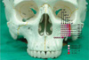
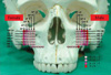
The horizontal base plane (HB) was defined as a plane perpendicular to the midsagittal plane and passing the most inferior border of the zygomatic process of the maxilla or zygomatic bone (Fig. 1A). Six more horizontal planes (HB-2, HB+2, HB+4, HB+6, HB+8, HB+10) parallel with the HB were constructed at 2-mm intervals. The bone located lower than HB-2 was not estimated in the present study because the developing tooth germ or root apex was observed in several patients with late mixed dentition or early permanent dentition.
On each horizontal plane of HB, HB+2, HB+4, HB+6, HB+8, and HB+10, the antero-posterior line (AP) was drawn parallel with the midsagittal plane and passing through the most anterior point of the infratemporal fossa (Fig. 1B, arrow). Five more antero-posterior lines (AP-2, AP-4, AP-6, AP-8, and AP-10) were constructed at 2-mm intervals. On the horizontal plane of HB-2, AP was defined as the line parallel with the midsagittal plane and passing through the most lateral point of the maxilla. The vertical lines of AP-2, AP-4, AP-6, AP-8, and AP-10 were also drawn on HB-2 at 2-mm intervals (Fig. 1A). The bone thickness was measured at the points where the anteroposterior lines and outer surface of the bone intersect at each horizontal plane. All of the measurements were performed by one investigator.
Method errors
The reliability of the measurements was determined on two arbitrarily selected samples. The bone thickness at each point was measured twice, separated by a one-week interval. The method error was calculated from Dahlberg's formula (method error=√∑d2/2n where d is the difference between 2 measurements of a pair, and n is the number of subjects). The method error of the measurement ranged from 0.06 mm to 0.30 mm with a mean of 0.19 mm.
where d is the difference between 2 measurements of a pair, and n is the number of subjects). The method error of the measurement ranged from 0.06 mm to 0.30 mm with a mean of 0.19 mm.Results
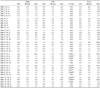
The minimum, median, and maximum values of the right and left sides and statistical difference are summarized in Table 1. Ten out of the 35 locations showed significant differences between the right and left sides, but the differences were less than 1.0 mm. The thickest point (HB+10, AP-2) was 5.0 mm on average, and the thinnest point was 1.1 mm. Figure 2 shows the color maps of the median values of bone thicknesses in the infrazygomatic crest and the zygomatic process of the maxilla. There was a tendency for the bone to be thicker at the superior and lateral area of the zygomatic process of the maxilla than the other locations.
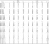
Table 2 shows the bone thickness in the male and female groups. Six out of 35 locations showed significant differences between the male and female groups. Generally, the bone thickness of the male group was greater than that of the female group. Figure 3 displays color maps of the median values of bone thickness in the male and female groups.
Discussion
In the present study, the FH plane was used as a horizontal plane from which to measure the bone thickness. For the consistent measurement of bone thickness, CT images were taken in parallel with the FH plane of the subjects. For the consistency and convenience of measurement, the binary image setting (window width: 0 HU, window level: 500 HU) was used. A pilot study had been carried out to determine the appropriate window level of the binary image setting. A CT had been taken with a cortical bone slice of bovine rib attached by periosteum. At the binary image of the window level of 500 HU, the bone thickness measured on the CT image was close to that measured at the cortical bone. Therefore, the binary image setting of the window level of 500 HU was used in this study.
The zygomaticomaxillary suture had been considered to be the reference area for constructing the horizontal planes and vertical lines; however, it was not clearly identified on the CT image in some children due to ossification. On the other hand, the most anterior point of infratemporal fossa was easily identifiable on the CT images on all of the horizontal planes, and these points were observed to be quite close to the zygomaticomaxillary suture.
The thickest point (5.0 mm) was at the point HB+10 and AP-2, which corresponded to the zygomatic process of the maxilla, and the thinnest point (1.1 mm) was at the point HB+2 and AP-10, which corresponded to the anterior wall of the maxillary sinus. The bone at AP-2 and AP-4 in all of the horizontal planes tended to be thicker than that at more medial points, and the bone thickness of these areas ranged from 1.8 mm to 5.0 mm, which was regarded as the appropriate thickness for the miniplate placement. Considering the thickness of the miniplate (2 mm), miniscrews of 4 to 7 mm in length can be selected. If oblique installation of the miniscrew is possible, a longer miniscrew could be selected for better bone support.
According to our findings, the bone thickness in the growing patients tended to be thicker at the superior and lateral areas of the zygomatic process of the maxilla. This might be related to the development of the maxillary sinus. In previous reports,17-20 the maxillary sinus continued to extend both laterally and inferiorly from the medial orbital wall after birth. Inferiorly, it reached the level of the hard palate at 9 years of age and continued to grow downward and together with the pneumatization of the maxillary alveolar bone, reaching the level of the nasal floor at 12 years of age. The floor of the maxillary sinus ended up extending 4-5 mm inferior to the nasal floor. The shape of the maxillary sinus also changed into a reverse pyramidal shape with the lateral expansion at the superior side.
Our results showed no differences between the right and left side except at 10 out of the 35 locations evaluated, and these differences were very small. The reason might be that the patients included in the present study had no facial asymmetry and the measurements were taken at symmetric locations. Previous studies have reported that the right and left side of the maxillary sinus showed no significant differences in the transverse width, anteroposterior width, height, or volume.17,18
Our results showed that only 6 out of 35 locations demonstrated a statistically significant gender difference. This is in concordance with many previous studies showing no significant difference in maxillary sinus development between males and females.17-20 However, there was a tendency for the bone to be thicker in males than females in the present study. This was probably related to the gender difference in occlusal forces because a high concentration of occlusal stress at the zygomatic arch might be related with thick and dense cortical bone of this area, with bony adaptation to the functional load.21
In some younger children with mixed dentition in the present study, the tooth germs or root apex of the permanent teeth were observed under HB-2. Therefore, the area superior to HB-2 seemed to be appropriate for placement of miniplates in children with mixed dentition. In a previous study, the distance from the apex of the mesio-buccal root of the maxillary second molar to the buccal bone plate was 4.63 mm.21 However, no study has evaluated the buccal bone thickness to the tooth germsl in children with mixed dentition. This should be investigated in a future study.
The patients in the present study were diagnosed with skeletal Class III malocclusion with maxillary hypoplasia. There might be differences in the bone thicknesses at the infrazygomatic crest area among skeletal Class I, II, and III patients. Further studies are needed to investigate the bone thickness with various skeletal patterns.
The principal conclusions can be summarized as follows. In the infrazygomatic crest area, the superior and lateral area of the zygomatic process of the maxilla are most appropriate for placement of a miniplate for orthodontic treatment in growing children with skeletal class III malocclusion and a retruded maxilla.
There was a tendency for the bone to be thicker at the superior and lateral area of the zygomatic process of the maxilla. There was no clinically significant difference in bone thickness between the right and left sides; however, the bone was thicker in the males than the females.

 XML Download