

 PDF
PDF ePub
ePub Citation
 Print
Print


Introduction
Recently, digital radiography has superseded film-based radiography because of its advantages. The lower diagnostic dose, no need for chemical development and fixation, the possibility of processing and analysis, image enhancement capacity, and image transfer are the main advantages of digital radiography.1-3 Several studies have shown that digital radiography has equal or better diagnostic accuracy compared to film-based radiography;4-12 therefore, digital radiography is widely used in dentistry, especially in endodontics.
Manipulation of images is one of the most important properties of digital radiography.13,14 Several digital processing algorithms are available that enhance the display quality of digital images.14,15 Since digital sensors produce a large amount of noise, different image processing schemes have been developed to improve image quality.16 Noise reduction with the use of different techniques might result in higher signal-to-noise ratios. For medical imaging, a high signal-to-noise ratio is considered essential for clinical diagnosis.16 Noise reduction, sharpening-smoothening, and edge enhancement are examples of these algorithms. It has been accepted that reduction of the noise value has a significant effect on the improvement of digital image quality. However, this process can lead to loss of some small structures on images.
Several studies have evaluated the use of various digital enhancement algorithms and their effects on diagnostic accuracy.17-25 However, there has been only limited research on the task-specific use of noise reduction algorithms.20,26-30 Some of these studies have shown that this type of enhancement does not change the diagnostic accuracy;20,29 on the other hand, others have shown the opposite.26-28,30
The effect of this enhancement on the measurement accuracy is particularly important in endodontics. The accuracy of measurement can influence the final verification of the outcome of root canal treatment by affecting the precise working length, and consequently, the potentially successful treatment. Therefore, the aim of the present study was to evaluate whether the length measurement of small endodontic files can be affected by application of noise reduction enhancement or not.
Materials and Methods
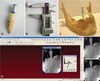
Thirty-five human-extracted single-rooted permanent teeth with intact crowns and roots were used in this experimental study. The teeth had only one root canal, measuring 20-24 mm in length. Periodontal or prosthetic problems were the reasons for extraction of the teeth. The samples were cleaned and disinfected by scaling and soaking in 0.5% sodium hypochlorite for 12 h and stored in distilled water at 4℃ during the study. Conventional periapical radiography was used to verify any abnormalities or pathologies, such as internal/external root resorption, root canal obliteration/calcification, or severe curvature. An anatomic access cavity was prepared with #008 and #010 fissure burs and an Endo-Z bur (Dentsply Maillefer, Ballaigues, Switzerland) in a high-speed handpiece. Gates-Glidden #3 and #4 drills (Dentsply Maillefer, Ballaigues, Switzerland) were used to enlarge the coronal and middle thirds of the root canals. ISO #08 K-files (Mani Inc., Utsunomiya, Japan) were inserted into the canals until the tips of the files were just visible at the apical foramen (Fig. 1A). The file length was measured with a digital caliper to the nearest 0.01 millimeter (Guilin Guanglu Measuring Instrument Co., Ltd., Guilin, China), and the file was shortened by 0.5 mm, which was set as the standard value for the endodontic file length (Fig. 1B). Only teeth with a length of 20-24 mm were included. Then, the files were inserted into the canals again and fixed with flowable composite resin (Tetric® Flow, Ivoclar Vivadent AG, Schaan, Liechtenstein). Afterward, the samples were placed in suitable sockets of a dry human mandible to reproduce the bone density.
In order to eliminate the radiographic magnification, a 10-mm round orthodontic wire was placed in the adjacent dental socket and fixed with wax. A Rinn-Endo-Ray film holder (Dentsply/Rinn Corporation, Elgin, IL, USA) was used to ensure parallelism. The standard geometric configuration was fixed at a 30-cm source-to-object distance. Radiographic images of each sample were obtained with Digora storage phosphor plates (Soredex Corp, Helsinki, Finland) and its special scanner, the Digora Optime (Soredex Corp, Helsinki, Finland), using a Prostyle dental X-ray unit (Planmeca OY, Helsinki, Finland) operating at 63 kVp, 8 mA, and 1.5-mm Al-equivalent filtration for 0.03 s. The digital images were numbered by Scanora 5.0 software (Soredex Corp, Helsinki, Finland) (Fig. 1D) in a semi-dark room and saved in the DICOM format for further processing and analysis (Fig. 1E). The noise of each image was reduced by single clicking on the "noise reduction" option on the diagnostic tools menu bar on the top of the operating window of the Scanora software, and the resultant image was saved again in the same format (Fig. 1F). Then, two blinded radiologists with 5 years or more experience in the interpretation of digital radiographs, identified the endodontic file tip and the most apical portion of the rubber stop of each file in the original and enhanced digital images. They also determined the most coronal and apical point of the orthodontic wire in the adjacent dental socket. The endodontic file and orthodontic wire lengths were measured by a third person using the measurement tool of the software to the nearest 0.1 mm. The magnification coefficient of each image was determined using real and radiographic orthodontic wire lengths. To eliminate the magnification effect of the radiography, the obtained endodontic file length for each image was divided by the magnification coefficient. Then, the mean values of the radiologist measurements were taken as data.
The data were first verified with the Kolmogorov-Smirnov test for the normality of data distribution. Repeated measures ANOVA and the Bonferroni test were used to compare the standard value and radiographic file length with and without enhancement. A SPSS software (ver. 11.0, SPSS Inc., Chicago, IL, USA) was used for analysis. Statistical significance was set at a confidence level of 95%. Also, Cohen's kappa statistic was used to assess interobserver reliability.
Results
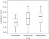
Repeated measures ANOVA showed significant differences between the standard, original, and enhanced images in the endodontic file length measurements (P<0.05). The Bonferroni test showed no significant differences between the two sets of digital radiographs, suggesting no improvement or reduction in the linear measurement accuracy with noise reduction digital enhancement (P>0.05); however, there was a tendency to overestimate the file length determination by digital enhancement. Additionally, there were significant differences between the two sets of digital radiographs and the standard value (P<0.05). Both the original and enhanced digital images had a tendency to overestimate the file length measurements (P<0.05). The results are summarized in Table 1 and Figure 2.
In addition, inter-observer agreement was excellent for the assessment of digital images with and without enhancement, showing a kappa value of 0.86.
Discussion
The main aim of digital processing is to create images with sufficient detail. This is possible by displaying information that has been collected during imaging but is not visible.19 Although processing should be able to improve diagnostic signals, some of the information might be lost during the process.18,19,23,31 Therefore, digital enhancement should be used with caution, based on the diagnostic task. This task-specific use of enhancements requires accurate investigation. The present study was the first one undertaken to evaluate the influence of noise reduction enhancement on the measurement accuracy of endodontic file length on images acquired using Digora storage phosphor plates. The accuracy was assessed by comparing the position of reproducible landmarks in the original and processed images.
The results of the present study showed that noise reduction does not decrease the accuracy of file length measurement. From the clinical viewpoint, this option does not result in the loss of subtle information, including the position of the tip of a #08 endodontic file. This result is consistent with the results reported by Koob et al20 and Haak and Wicht.29 They showed that noise reduction does not change the diagnostic quality of images in identification of interproximal caries. In addition, Koob et al20 reported that there were no significant differences between images with and images without noise reduction in determination of interproximal caries depth. In contrast, in three studies,26-28 Davies demonstrated that the use of noise elimination filters resulted in size and shape distortions. Furthermore, Xu and Lai30 reported that these distortions occurred in fine details such as the tip of endodontic files, particularly with #06 and #08 files. In addition, Brullmann et al16,32 evaluated noise reduction in two different situations. In the first study,16 it was illustrated that noise filtering had no effect on the accuracy of length measurement of files greater than #10, but this enhancement resulted in underestimation of the length of #10 and smaller endodontic files. This is in contrast with the result of the present study showing overestimation in the length measurement. This controversy may be due to differences in the study design such as the use of charge-coupled device (CCD) digital radiography rather than a PSP system, the use of digital software (Sidexis XG 2.4, Sirona, Bensheim, Germany) with a special filtering program (Borland C-Builder 6.0, Borland GmbH, Langen, Germany) rather than Scanora software, the presence of a marker in the apical portion of the root of the teeth that helps in the identification of the tip of the endodontic file, and the existence of the soft tissue of the mandible resulting in more scatter radiation and noise in that study. In the second study,32 it was shown that this filter was effective in the elimination of noise without the loss of diagnostic information but did not increase the odds of identification of dental root fracture.
Some studies have addressed subjective image quality with and without noise reduction based on observer performance.29,33,34 Their major limitation was reliance on subjective assessment of images. For example, Yalcinkaya et al33 compared the display quality of anatomical structures on conventional and digital periapical and panoramic radiographs. They reported that conventional radiography was superior to unfiltered and noise-reduced filtered digital images, with no statistically significant differences between filtered and unfiltered digital images. However, it has been suggested that it may be more important to use a computable objective measure to predict diagnostic accuracy rather than subjective assessments. Näslund et al34 reported on both objective and subjective evaluations of image quality with and without post-processing noise reduction. The authors basically compared the ability of localization of anatomic landmarks on standard exposed, low-exposed, and a combination of low-exposed and noise-reduced digital cephalograms. They reported that the landmarks were identified more effectively on the low-exposed images than on the images with post-processing noise reduction, although the subjective evaluation of the image quality indicated the opposite. The standard-exposed digital cephalograms were the best in objective and subjective quality evaluations. The results of the objective evaluation contrasted with the result of the present study.
In addition, the results of the present study showed that both enhanced and unenhanced digital images tended to overestimate the length of endodontic files, consistent with the results of studies by Mehdizadeh et al35 and Williams et al.36 In contrast, Schmitd et al37 and Brito-Junior et al38 reported that the measurement accuracy of digital images was comparable to real measurements.
It is undisputed that the diagnostic impact of digital imaging depends on the task, the quality of source data, and the kind of image processing applied.19 Therefore, in a clinical situation, discrimination of small file tips may become more difficult. Selection of optimal exposure time is important because under-exposed images have a higher amount of noise.39 In addition, the presence of soft tissues and the increase in the volume and density of hard tissues in the clinical situation results in a greater amount of scattered radiation, increasing the amount of noise. Therefore, clinical studies are suggested for further evaluation of the usefulness of noise reduction digital enhancement in practice.
In addition, although a storage phosphor plate system was used in the present study, most endodontists benefit from the advantages of solid-state detectors to obtain periapical radiographs during root canal treatment. Therefore, the application of solid-state detectors is also recommended for further investigation.
In conclusion, noise reduction enhancement did not change the measurement accuracy of endodontic file length on digital images. Therefore, it could be used, depending on the demands of the practitioner.

 XML Download