

 PDF
PDF ePub
ePub Citation
 Print
Print


Introduction
Although the diagnostic information provided by radiographs may be of definite benefit to the patient, the radiographic examination carries the potential for harm from ionizing radiation inducing carcinogenesis. A statistical association between X-ray exposures in dentistry and increased incidence of salivary gland tumors,1 thyroid cancer,2 and intracranial meningioma3 has been reported.
Although the radiation doses used by dentists might be low for individual examinations, patients are exposed to repeated examinations over time, and many people are exposed during the course of dental care. The unregulated habit of taking of dental radiographs based on a single frequency for all patients could lead to unnecessary patient exposure.4
The justification and optimization of radiography is now an important issue for dental practitioners in terms of reducing radiation dose. The selection criteria for radiographs in dentistry has been revised recently in accordance with guidelines and peer-reviewed research materials of USA and Europe,5-9 but little has been published on this subject in Korea. To date, many types of equipment and techniques have been developed in dental radiography to reduce patients' exposure dose.10 Good radiologic examination practice includes appropriate collimation, use of a lead apron and thyroid collar as well as application of objective selection criteria.11
There has been no internationally published data about the attitudes of dentists in Korea regarding radiation safety regulations. The aim of this study was to survey the attitude of Korean dentists regarding radiation safety and use of dental radiography selection criteria.
Materials and Methods
A total of 267 Korean dentists participated in this study. Among them, 67 dentists were recruited from the attendees of the 2012 Fall Science Meeting of Korean Academy of Oral and Maxillofacial Radiology. Two hundred additional dentists participated through a web-based survey system. Five questions were asked about radiation protection of patients and operators during dental radiograph examinations. The topics included: 1. Prescriptions of dental radiographs A) routine examinations, "routine" means the predetermined initial radiographic examination on new patients seeking comprehensive care. The example of routine is a panoramic radiograph as part of screening process for occult disease, or the full-mouth periapical+ bitewing+ panoramic radiographies combination regardless of signs and symptoms. B) selective radiographic examinations based upon the patient's clinical signs, symptoms, and history using selection criteria,12 C) imaging based on the 2004 Food and Drug Administration-American Dental Association (FDA-ADA) guideline compliance,13 D) the frequency of bitewing radiography. 2. The frequency of communication with patients 3. Intraoral image receptor use A) type of image receptor (digital sensor/film) B) placement of image receptor in patient's mouth. 4. The use of lead apron/thyroid collar 5. The use of rectangular collimation. Among these factors, image receptor placement was the factor closely related to operator protection. The detailed questions were as follows.
1. Prescription of dental radiographs
A) Do you routinely prescribe radiographic examinations or choose to make selective radiography based on any guideline when new patients come to your dental clinic?
B) Do you regularly use bitewing radiographs?
2. Communication with patients
A) How often do you encounter patient questions about radiation safety?
B) How often do you explain radiation risk/benefit to patients and acquire patient consent before taking radiographs?
3. Image receptor
A) Which receptor do you use among film, digital sensor, and photostimulable phosphor (PSP) plate?
B) Regarding the image receptor placement, who usually holds the film/digital receptor in patient's mouth during the intraoral X-ray exposure?
4. The use of lead apron or thyroid collars
Do you always cover patients with lead apron or thyroid collar during radiographic examinations?
5. Rectangular collimation
Do you regularly use a rectangular collimator during intraoral radiographic examinations?
The respondents were classified into two groups including general dentists (n=240) and specialists of oral and maxillofacial radiology (n=27, formally trained or being trained by residency program). The purpose of this classification was to determine whether the specialists were more informed about radiation safety.
Results
Prescription of dental radiographs
Dentists utilized the predetermined routine examination (34.1%), selective radiography (64.0%), and guideline compliance (1.9%). While 36.8% of the general dentists expressed a preference for routine ordering of radiographs, only 12.1% of the specialists did (Table 1). Routine bitewing radiography was used by 35% of all of the dentists. Although 65.4% of the specialists preferred to take bitewing radiography only 31.2% of the general dentists had this preference (Table 2).
The frequency of communication with patients
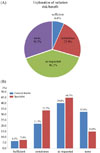
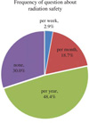
Patients asked about radiation hazards once or twice a month for 18.7% of respondents while patient inquiries were once or twice a year for 48.4% of the respondents. Patients asked no questions about radiation hazards for 30% of the practitioners (Fig. 1). "Occasional" explanation of radiation risk/benefit to patients was provided by 63.2% of the dentists; however, 30.2% of the dentists never explained radiation risk/benefit to patients (Fig. 2A). Of the dentists who never discussed radiation risk with patients 14.8% were the specialists compared with 32.0% of the general dentists (Fig. 2B).
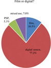
Intraoral image receptor
X-ray film was used by 10.5% of the dentists while digital sensors were used by 77.2% of the dentists (Fig. 3). Receptors were held by patients or guardians for 40% of the dentists, but 60% of the operators held the image receptor themselves during intraoral radiographic examinations. Patients or guardians held the image receptor during intraoral radiographic examinations for 69.2% of the specia-lists compared with 43% of the general dentists (Table 2).
The use of lead apron/thyroid collar
Lead apron/thyroid collars were used for patients in 21.7% of the dental offices. Two thirds of the specialists utilized apron/collar shielding; however, only 16.9% of the general dentists followed this practice (Table 2).
Discussion
Radiation protection of patients in dental radiology is related to the following factors: the appropriate selection criteria, equipment and film, and quality assurance programs.11 The most significant dose-associated factor is patient selection criteria. Patients who are likely to benefit from a particular radiographic examination are identified according to the selection criteria based on patient signs, symptoms, and history.14 In the 1950's, most dental schools in the United States (US) took full mouth intra-oral radiographic examinations as a diagnostic aid and screening tool.6 The International Commission on Radiological Protection (ICRP) then began to develop the risk/benefit concept. This concept suggested that all intentional exposure to a patient must be justified and be kept as low as possible.15 The American Dental Association (ADA) recently emphasized the necessity of using a specific type and frequency of radiographic examination over the previous concept of radiographic examination as a routine procedure.16 However, still, "As Low As Reasonably Achievable, ALARA)" principles are not strictly applied in the dental field,12 Many US and Canadian dental schools continue to obtain radiographic examinations routinely with new patients before a clinical exam.17 Among Canadian dental schools, the generalized use of routine radiography was common for dentate adult patients and the use of selective radiography was uncommon.12
In the present study, one third of Korean dentists made predetermined routine radiographic examinations while selective radiography was performed by 64.0% and FDA-ADA selection criteria by 1.9%. This result suggested that routine radiographic examination was relatively common and selection criteria guideline compliance was very low. It seems that little attention has been paid to radiographic selection criteria by dentists in Korea. Other type of radiographic examination such as panoramic radiography might also be carried out more frequently than needed. Routine radiographic examinations might increase patient exposure unnecessarily. Dentists could reduce radiation exposure by adopting patient selection guidelines.18,19 Radiographic examination should be decided according to the professional judgment of the dentist, taking into considering the type, frequency, and extent of the area to be examined.20 Periodic evaluation and revision, when indicated, of selection criteria guidelines is recommended for the safe use of dental radiography in Korea.
In this study, 30.2% of of respondents had not experienced patients' asking about radiation safety at all. While 32.0% of the general dentists had no experience explaining radiation risk/benefit to patients, even 14.8% of specialists had no such experience. Dentists should explain the treatment plan to the patient so that the patients can be involved in treatment decisions.21 Patients should be informed of possible untoward results of the treatment and have the right to refuse the proposed intervention.22 Patients have the right to take part in clinical decision making after being informed of significant risks, benefits, and alternatives.23
Bitewing radiography is well known for high detectability of interproximal caries24 and is also useful for evaluating the periodontal condition.25 However, the results of the present study showed that only 35% of respondents performed bitewing radiography limitedly. Korean dentists might be unaware of the value and recommended frequency of bitewing radiographs.
Many Korean dentists (77.2%) were using digital sensors despite the high cost of this type of equipment. The fact that many Korean dentists used digital intraoral sensors without receptor holders might be related to the relatively bulky thickness of digital sensors as compared to intraoral film. In addition, dentists who wanted to reduce the re-taking of radiographs due to a positioning error might hold the image receptor despite the radiation hazard. The present study showed that 60% of operators held the image receptor themselves during intraoral radiographic examinations. Operators should not hold the image receptor during exposure. The preferred method for operator protection would be to position the receptor with a holding device and to stand behind a protective barrier or outside the room during radiographic exposure.
Lead aprons and thyroid shields are patient-protective equipment that minimizes exposure from scattered radiation. However, protection of the abdomen has little relevance to dental radiation protection since scattered radiation to the gonadal area is small that the risk of hereditary effects is negligible.26 The use of a lead apron on patients may not be required if National Council for Radiation Protection & Measurements (NCRP) recommendations are followed strictly.27 The risk of long-term health effects from low dose exposure is uncertain.28 There has been some evidences that radiation exposure to the thyroid during pregnancy is associated with low birth weight.29 Protective thyroid collars substantially reduce radiation exposure to the thyroid during dental radiographic procedures.30 The present study showed that only 21.7% of dentists draped lead apron/thyroid collar over patients, indicating that a relatively high percentage of dentists were unaware of the potential of thyroid exposure or the possibility of minimizing this with the use of a thyroid collar/shield. Since every reasonable precaution should be taken to minimize radiation exposure, protective thyroid collars and aprons should be used whenever possible. This is recommended for all patients, especially for children, women of childbearing age, and pregnant women.13
The field size is one of the most important factors in dose reduction and the frequency of retakes.31 Restricting the X-ray beam to only the receptor area can prevent unnecessary patient exposure. By replacing round collimation with rectangular collimation, the effective dose would be reduced by almost 60%.32 Rectangular collimation did not significantly affect the diagnostic yield of radiographs despite the increase in minor cone cuts.33 The present study showed that 14.6% of Korean dentists used rectangular collimation. The percentage of dentists using rectangular collimators varied by country including Sweden (29%),34 Belgium (6%),35 Turkey (5.5%),10 and Canada (8%).36 Dentists can create rectangular beams by inserting a rectangular windowed piece of metal into the round position-indicating device (PID).37 Rectangular collimation can also be achieved with either a rectangular positioning-indicating device, or a rectangular collimator clipped to the aiming ring. Unfortunately, some Korean dentists might believe that collimation is inherent components of the tube housing of an X-ray machine. This misunderstanding could be related to the low rate of use of rectangular collimation of 14.6%.
Previous studies have reported differences between specialists and general dentists with regard to the use of rectangular collimation38 and selection criteria.17 The results of the current study suggested that specialists were more knowledgeable about selection criteria and radiation safety; however, even specialists showed little interest in explaining radiation risks and benefits to patients, utilizing bitewing technique, or applying rectangular collimation.
The majority of Korean dentists in this study did not currently follow the good radiological practices including the application of selection criteria, explanation of radiation risk and benefit, proper image receptor holding, and use of rectangular collimation. This study might provide the foundation for more detailed studies that would assess the attitudes toward radiation safety and quality assurance. Continuing education on radiation safety would be required for dental health care providers. Also, the development of Korean dental radiography selection criteria would be also recommended.


 XML Download