

 PDF
PDF ePub
ePub Citation
 Print
Print


Introduction
Injury to the inferior alveolar nerve bundle is a serious complication following the extraction of impacted mandibular third molars. Most reports have demonstrated an incidence range from 0.5% to 8%.1 In order to avoid inferior alveolar nerve injury, it is critical to evaluate the proximity of the mandibular canal to the third molar preoperatively. In the literature, several signs have been defined on panoramic radiographs, which indicate the close proximity of the mandibular canal to the third molar. Notably, four radiographic signs - darkening of the root, interruption of the cortical lines, diversion of the mandibular canal, and narrowing of the root - are recognized to be associated with inferior alveolar nerve proximity to the mandibular third molar on panoramic radiography.2
Although the incidence of this proximity is low, the complications can be serious, justifying more precise preoperative evaluation.3 Several aspects of the clinical usefulness of cone beam computed tomography (CBCT) have been discussed.4,5 However, patients are sometimes unnecessarily exposed to radiation even though conventional evaluation techniques are of high diagnostic value. Therefore, this study was designed to assess the reliability of panoramic radiographs in detecting radiographic signs of inferior alveolar nerve proximity evidenced on CBCT images.
Materials and Methods
This cross-sectional study was performed using panoramic and CBCT images of 96 patients referred by maxillofacial surgeons whose panoramic features suggested a close relationship between the inferior alveolar canal (IAC) and third molar root. The approval of the Ethical Board of the Institutional Ethics Committee of Shiraz University of Medical Sciences was obtained before conducting this study.
One hundred and thirty-two impacted mandibular third molars were included in the study. The patient group consisted of 50 females and 46 males with a mean age of 25.41 years. The panoramic radiographs were taken using a Proline XC unit (Planmeca, Helsinki, Finland) using a 15 cm×30 cm photostimulable phosphor receptor (Regius 110, Konica Minolta Medical & Graphic, Inc., Tokyo, Japan). The CBCT images were obtained with a Kodak 9000 (Carestream Health, Inc., Rochester, NY, USA) at 90 kVp, 6 mA, and 10 seconds. A maxillofacial radiologist with more than ten years of experience evaluated the panoramic and CBCT images independently to determine the topographic relationship between the impacted third molar and the mandibular canal.
For the CBCT images, the presence or absence of direct contact between the tooth root and the canal contents was three-dimensionally evaluated, which was used as the diagnostic criterion in predicting neurovascular bundle exposure. Direct contact was considered to be present when loss of canal cortical bone between the two structures was observed on axial, panoramic, and cross-sectional images. Moreover, any root dilacerations, buccal or lingual cortex perforation, and canal diversion were noted for each case.
The presence or absence of direct contact on the panoramic radiographs was evaluated by the following four features, all of which have been reported to be suggestive of close contact between the tooth root and the mandibular canal: (a) interruption of the mandibular canal wall, (b) darkening of the root, (c) diversion of the mandibular canal, and (d) root dilaceration.
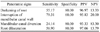
The sensitivity, specificity, positive predictive value (PPV), and negative predictive value (NPV) of each panoramic feature in depicting the association of the root on CBCT images were calculated. The diagnostic criterion for panoramic images was defined using logistic regression analysis to determine the independent predictive value of the four panoramic features listed in the aforementioned paragraph.
Results
The study sample consisted of 132 impacted third molars from 96 patients (50 females and 46 males). The most frequent finding by CBCT was a lingual course of the mandibular canal.
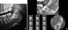
Contact of the tooth root with the canal was observed in all cases in which a loss of the cortical line of the canal or darkening of the roots was found on the panoramic radiographs (Fig. 1). Loss of the cortical line in the panoramic signs showed the highest sensitivity and was the most frequent radiographic sign for predicting association (Table 1).
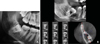
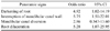
When darkening of the root was seen on a panoramic view, the risk of association in CBCT images was 4.92 times more (Table 2, Fig. 2). However, when darkening of root was detected on panoramic view, the risk of cortex perforation in CBCT images was just 2.8 times greater (CI=1.18-6.62). When an interrupted border of the mandibular canal was detected on panoramic radiography, the risk of association in CBCT images was 5.75 times greater. Root dilacerations on panoramic radiography raised the chance of association in CBCT images to 5.28 times greater (Table 2).
Discussion
Although CBCT provides a better evaluation of anatomical structures and greater intra-operative safety, panoramic radiography is still the most widely used technique for assessing the relationship of the third molar root and canal. Several clinical studies have determined the specific radiographic signs detected on panoramic radiographs to be suggestive of a close relationship between the third molars and mandibular canal.2,6,8,11
The results of this study showed that four radiographic signs of proximity on panoramic radiography could be used to predict the association of the canal and root although each was of different value.
Interruption of the cortical lines on a panoramic image raised the risk of root and canal contact on CBCT images (odds ratio=5.75) and showed the highest sensitivity among the four signs of association. This result was in accordance with the studies by Nakagawa et al1 and Jung et al6 and indicated that the risk of contact between the mandibular canal and the third molar root was enhanced when the white line of the canal was not visible on panoramic radiographs.
Darkening of the third molar roots where the mandibular canal was superimposed has been reported to be suggestive of an intimate relationship between the root and canal in several studies.7-10 Some others believed that this radiographic sign indicated thinning or perforation of the lingual cortical plate.6,11 The results of the present study showed that the risk of the third molar root contact with the canal in the cases wherein the panoramic sign of the darkening of the roots was observed was higher than in cases of lingual cortical plate perforation (odds ratio=4.92 and 2.86, respectively). In contrast to the results of Tantanapornkul et al11 and Jung et al,6 our study sample consisted mostly of cases in which the canal was located lingual to the roots. This might explain the reason that less lingual cortical plate perforation was detected in the present study. However, it also emphasizes the importance of using an advanced imaging technique to explore whether darkening of the third molar root is related to canal contact or cortical plate perforation.
The probability of molar root association with the mandibular canal in the presence of related radiographic signs, known as positive predictive value (PPV), was highest for dilaceration and darkening of the roots in our study. The negative predictive value (NPV) is the probability of no root association with the canal when these radiographic signs are absent; and it was low for all signs except for canal diversion.
Mandibular canal diversion had the least sensitivity to predicting association, which was in contrast with previously published studies.3,8,12 This difference could be due to patient selection in our study. Since the referred patients were suspected of a relationship between the canal and root on panoramic radiography, cases which had a sign of a change in the path of the canal might have been less likely to be referred. However, our findings were in agreement with the study of Neves et al13 in which mandibular canal diversion had lower importance in predicting the association of the canal and root.
In the present study, root dilaceration showed a higher sensitivity than mandibular canal diversion in predicting the association of the canal and root. This finding suggested the possibility of root displacement during the maturation of adjacent anatomic structures rather than the alveolar canal. Detecting root dilaceration might affect surgical planning for the mandibular third molars.
Although the absence of cortical bone in CBCT is highly suggestive of nerve exposure, which has been related with an increased risk of nerve damage, reports indicate a 20% risk of postsurgical paresthesia, with a 70% chance of recovery by one year after surgery.14 This recovery rate showed that anatomical contact was not the sole determining factor for postsurgical prognosis. The association of the inferior alveolar nerve with the root could be clearly visualized using magnetic resonance imaging.15 Further studies are recommended to assess to what extent radiographic findings of nerve proximity can predict postsurgical complications. However, patients with a detected association of the canal to the root should be considered at high risk of nerve damage.
 XML Download