

 PDF
PDF ePub
ePub Citation
 Print
Print


Introduction
Biological and chronological ages are not completely associated with body development. Therefore, parameters are obtained to evaluate biological development such as dental age, bone age, mental age, and other factors such as menarche, voice change, height, and weight.1 Dental age assessment plays an important role in forensic medicine, pediatric dentistry, and orthodontic treatment planning.2 Several methods of age estimation have been suggested, and the method by Demirjian et al3 is the most frequently used.4,5
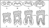
Demirjian's method is based on eight stages of tooth calcification ranging from crown and root formation to apex closure of the seven left permanent mandibular teeth. The score of each stage is allocated, and the sum of the scores gives an evaluation of the subject's dental maturity. These scores can be converted into a dental age by means of available tables and percentile curves from the original study. The difference between the dental and chronological age indicates an advancement or delay in dental maturity.6
Estimating dental maturity according to percentile charts has been shown to be more suitable in determining whether the dental maturity of a subject with a known age is advanced or delayed rather than to predict an unknown age.4,7,8 Demirjian's study was conducted from the data obtained from a group of French Canadian boys and girls. Thereafter, several studies have shown that the estimation of dental age of another population using Demirjian's standards developed for French Canadians was not suitable for their study populations and the ethnic differences of dental maturity had to be taken into account.9-13
The establishment of population-specific standards for third molars is considered important for enhancing the accuracy of age estimation based on the development of these teeth. However, third molars are often missing congenitally or have been extracted, so they cannot be depended upon to indicate age. Moreover, if they are malformed or malpositioned, age estimation may be difficult.7,14 To overcome these limitations, the second molars can be analyzed as an age indicator. Many studies have investigated the accuracy of forensic age estimation based on the chronology of second molar development and have concluded that it can be considered a valuable age indicator in populations.7,14,15
Thevissen et al16 insisted that dental age estimation, especially for young individuals, should be based on the data collected from the appropriate population group. Thus, the aim of this study was to evaluate the chronology of second molar development in Brazilians and suggest its application to forensic age estimation.
Materials and Methods
This study was approved by the Center for Health Sciences Ethics Committee in Human Research of Federal University of Paraíba, Brazil (0178/2009). A cross-sectional analysis was performed for a sample of 457 panoramic radiographs taken from 192 males and 265 females aged between 4.6 and 16 years. A convenience sampling method was used, and the panoramic radiographs had been obtained from July to December 2010 from a private dental radiology service in Paraíba, Brazil. A sample of 457 out of a total of 1,854 panoramic radiographs was selected based on the following criteria: the analysis was restricted to individuals of both genders aged 3-16 years and born in the state of Paraíba, Brazil. Unreadable radiographs, or those showing hypodontia or gross pathologic problems, were excluded.
The panoramic radiographs were taken using a Rotograph Plus panoramic machine (Villa Sistemi Medicali, Milan, Italy), and suitable exposure factors considering the patients' ages and sizes were applied. The Kodak T-Mat G/RA X-ray film (Eastman Kodak, Rochester, NY, USA) were manually processed according to temperature/time tables using fresh Kodak concentrated processing solutions (Eastman Kodak, Rochester, NY, USA). The radiographs were digitized using a Hewlett-Packard scanner (Hewlett-Packard Co., Palo Alto, CA, USA) equipped with a transparency reader as part of routine clinical activity, with a resolution of 300 dots per inch (dpi). All the images were saved and stored in Tagged Image File Format (TIFF). Each digitized panoramic image was coded with a numerical ID. The examinations were assessed twice by a radiologist with 5 years of experience. Then, all of the images were exported to the computer and viewed, randomly, in a quiet and darkened room with the aid of a 17-inch LCD monitor (HP Compaq LA1751 g, Hewlett-Packard Co., Palo Alto, CA, USA) using the Windows Picture and Fax Viewer (Microsoft, Redmond, WA, USA).
The age and gender of the subjects were thus unknown to the observer. Prior to the examination, verbal and practical instructions and calibration tests were performed. The evaluation method was explained, and training and knowledge of the evaluators were verified. A minimum interval of 30 days between the evaluations of the same patient was established. If the first and second assessments differed, the readings were repeated until a consistent result was obtained. All of the second molars were assessed. For the analysis of the degree of calcification, Demirjian's method (Fig. 1) and a form was filled out for each panoramic radiograph were used. The examiner could use the zoom tool whenever necessary to obtain a maximum magnification of 3 times to avoid loss of resolution. At most, 10 panoramic images were examined each day to avoid compromising the assessments due to eyestrain.
The data were analyzed using Stata® 9.2 (StataCorp, College Station, TX, USA) and Minitab® 15 (Minitab Inc., State College, MA, USA) software. To evaluate the relationship between age, the calcification level proposed by Demirjian, gender, and tooth, a multiple linear regression model was adjusted taking age into consideration as the response variable. In all the tests, a 0.05 significance level was adopted.
Results
The patients' ages ranged from 4.66 to 16 years (11.7±2.5 years). A total of 192 (42%) panoramic radiographs were taken from male individuals, and 265 (58%) were taken from female individuals.
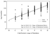
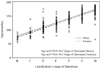
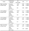
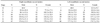
Table 1 presents the results of the multilinear regression models adjusted for each second molar to predict the age (in months) of patients in the sample based on the gender and Demirjian's score for each second molar. There was a statistically significant difference between the average ages of the patients in the different stages of calcification of second molars according to the gender (p<0.05). The results of the regression models corresponding to the right maxillary second molar and left maxillary second molar were found to be identical. The same was observed for the left mandibular second molar and right mandibular second molar. There were no second molars in the sample of patients in stage A of calcification; thus, the first stage of calcification found was stage B (B coefficient). For the upper second molar teeth, stage B had a mean age of 76.6 months, and the lower ones, an average of 65.5 months old. In the upper teeth, there was a variation of 16.1 months for the subsequent stage and in the lower teeth, an interval of 18.1 months.
Figures 2 and 3 present the results of the multilinear regression models adjusted to predict the age of patients in the sample based on gender and the second upper and lower molars, respectively. There was a significant association between age and Demirjian's calcification stages of the second upper and lower molars. A similar situation is observed when gender is the variable compared. In each stage, the average age of the male patients was higher than that of the female patients. These values make possible the development of formulas to predict the patient's age based on the calcification stage of the second molar evaluated.
Discussion
Estimation of age at death of human remains constitutes an important contribution to the identification of missing people, and also provides valuable data in the paleodemographic analysis of historical skeletal remains.17 In this study, based on the results, it was possible to develop formulas to predict chronological age in a specific Brazilian population, contributing significantly to age estimation in forensic evaluations.
Schmeling et al18 noted the influence of ethnicity on the development of systems of age estimation and the difficulty when the subjects of forensic examination mostly belonged to populations for which no reference studies could be available for forensic purposes. Following this line of thought, Chen et al6 observed that the development standards used by Demirjian et al for French-Canadian children were not suitable for western Chinese children of different origins; therefore, it is necessary to establish new tables for these populations.
In the present study, the method proposed by Demirjian et al was chosen as the assessment parameter to be compared to the international literature. In Brazil, the method of Nolla19 was frequently adopted. Moreover, we considered the need to evaluate dental age in the Brazilian population. In fact, there has been significantly less research on genetic, environmental, and even nutritional characteristics of the Brazilian population compared to other populations.
External influences, such as some environmental factors, socio-economic status, nutrition, dietary habits, and lifestyle, despite having little impact on dental development, should be taken into consideration.2 Phillips and van Wyk Kotze12 suggested that the socio-economic environment of a child might play a role in the calcification of the permanent teeth and influence the radiographic images of these teeth for aging purposes. This would explain why the pubertal growth spurt did not occur at the same time for all individuals. The sample used in the present study was not evaluated for external and internal modifying factors. However, the participants probably had a high socioeconomic status since they were the patients of a private clinic.
Some studies have suggested restricting the evaluation of dental development to the lower elements because maxillary teeth cannot be easily seen on panoramic radiographs and little data may be available for these teeth.8,11 Nevertheless, in the present study, the sample was selected based on the possibility of detailed visualization of the degrees of calcification of the second molars. Therefore, 10 unclear panoramic images of the upper molars were excluded.
It has been postulated that Demirjian's dataset is only applicable for individuals aged from 3 years to 16 years.9 Furthermore, the minimum age of patients in the present study was established based on Moyers,20 who explained that the development of permanent second molars began at 3 years of age for both genders. Although a minimum age of 3 and maximum of 16 years was a sample selection criterion in the present study, there were no patients aged less than 4 years and 9 months. This could be explained by the need for a solid justification for taking panoramic radiographs before the mixed dentition period. Furthermore, younger children were usually unable to stay perfectly still for 15 to 20 seconds, which might lead to incorrect patient positioning and distortion of the panoramic radiograph.
In the present study, there was a correlation with chronological age. This result was similar to the findings of others.4,5,7,14,15,21
According to the study by Bagherian and Sadeghi,5 dental development, including that of the permanent second molars, was earlier in females than in males. This fact was also observed in the present study, with females showing a precocity of 4.3 to 4.4 months for the upper teeth and 3.0 to 3.7 months for the lower ones. However, Nolla did not find significant differences in the degree of mineralization of teeth between the genders.19
Nolla19 noticed that neither the upper nor the lower teeth in either gender showed significant differences in the degree of mineralization on the right and left side. On the other hand, in the present study, the degree of calcification was significantly different between the lower and upper teeth, with the former growing earlier than the latter, which was consistent with the findings of the study by Holtgrave et al22 carried out in the United Kingdom.
Our results indicated that the dental development in females could be considered to occur earlier than in males; in addition, the lower second molar teeth develop much earlier than the upper ones, and there was no difference between the right and left second permanent molars. In conclusion, it was observed that ethnic variables were related to certain parameters of age and gender in a Brazilian population, contributing important information for forensic evaluations.

 XML Download