

 PDF
PDF ePub
ePub Citation
 Print
Print


Introduction
The maxillary sinus varies in its extension. It is essential to understand the anatomic relationship between the maxillary sinus floor and the root of the maxillary molar for planning preoperative treatments for maxillary posterior teeth.1
The close relationship of the maxillary sinus and the roots of the maxillary molars can lead to accidental oroantral communication.2,3 The topographical relationship of the roots of the posterior maxillary teeth and the maxillary sinus floor is an important determinant in the prognosis of orthodontic tooth movement.4 Sinusitis can result from the spread of a periapical or periodontal infection to the sinus or iatrogenic perforation of the sinus floor.3,5
It has been reported that the anatomical relationship between the root and the cortical plates might influence the spread of odontogenic infection originating in the maxillary molars.6 The thickness of the bone between the root and the alveolar cortical plate might be important in predicting the spread of infection as well as in treatment planning.
The purposes of this study were to investigate the relationship between the roots of the maxillary molars and the maxillary sinus using cone beam computed tomography (CBCT) and to measure the distances between the roots of the maxillary molars and sinus floor and the thickness of the bone between the root and the alveolar cortical plate.
Materials and Methods
The study samples were selected randomly from the patients who had visited Pusan National University Hospital between 2010 and 2011 and had normally erupted bilateral maxillary first and second molars, as shown on the panoramic radiographs. The selected patients underwent CBCT examination due to pathology such as cysts, tumors, impacted third molars, or temporomandibular joint disorders. Subjects with pathology in the maxillary posterior teeth were excluded from the study. A total of 332 maxillary molars in 83 patients were examined using CBCT images. The sample of patients comprised 33 males and 50 females with a mean age of 28.8 years, ranging from 20 to 53 years.
CBCT images were acquired with a CBCT scanner (PaX-Zenith 3D, Vatech Co., Hwaseong, Korea). Scanning parameters were 110 kVp, 24 seconds, 5.7 mA, a voxel size of 0.2 mm or 0.3 mm, and a field of view of 16cm×14 cm or 24 cm×19 cm. The CBCT volume data were reconstructed using CBCT software (Ez3D2009, Vatech Co., Hwaseong, Korea). CBCT images were evaluated to assess the roots of the maxillary molars, maxillary sinus, and cortical plate.
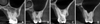
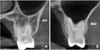
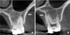
The vertical relationship between each root of the molar and the sinus floor was classified into four types based on the CBCT cross-sectional images: Type 0, the root was not in contact with the cortical borders of the sinus; Type 1, the root was in contact with the cortical borders of the sinus; Type 2, the root was projecting laterally on the sinus cavity, but its apex was outside the sinus borders; and Type 3, the root apex was projecting into the sinus cavity (Fig. 1).7 In Types 2 and 3, the horizontal relationship between the roots of the teeth and the sinus floor was classified into three types: Type B, the lowest point of the sinus floor was located on the buccal side; Type BP, the lowest point of the sinus floor was located between the buccal and palatal roots; Type P, the lowest point of the sinus floor was located on the palatal side of the palatal root (Fig. 2).7 In Type 0 and Type 3, the distance between the apices of the molars and the sinus floor was measured using CBCT cross-sectional images. The measurements were taken from the root apex to the cortical inferior wall of the sinus along the longitudinal axis of the root. The apices extending below the sinus floor were assigned positive values, whereas those above the sinus floor were assigned negative values (Fig. 3). The minimum distance from the root below bifurcation to the appropriate cortical plate was measured in the CBCT cross-sectional images. For buccal roots, the distance to the buccal cortical plate was obtained, and the distance to the palatal cortical plate was obtained for the palatal roots (Fig. 4).
Intra-observer variation was determined by repeating all of the measurements at an interval of four weeks. No statistical differences were found using a paired t-test between the replicate measurements. The averages of two measurements were then computed. A paired t-test was used to compare the measurements of the left and right sides. The results from the right and left molars were averaged for each tooth type because no statistically significant differences were found between the right and left side measurements. A descriptive analysis of the data was presented as frequency, mean, and standard deviation (SD). A one-way analysis of variance (ANOVA) test was used to assess the differences of bone thickness covering the roots among four vertical relationships between the root and the sinus. A p-value under 0.01 was considered statistically significant. All of the statistical analyses were carried out with statistical software (PASW Statistics 18.0, SPSS Inc., Chicago, IL, USA).
Results
In the classification of the each root in relationship to the sinus floor, Type 3 was more frequent in the buccal roots, including the mesiobuccal and distobuccal roots, of the maxillary molars. Type 2 was most common in the palatal roots of the maxillary first molar (M1), whereas Type 0 was most frequently observed in the palatal roots of the maxillary second molar (M2) (Table 1). The vertical relationship was classified in each molar, and Type 3 was shown to be most frequent in the molars with more than one root (Table 2). In the horizontal relationship between the roots of molars and the sinus floor, Type BP was most frequent in molars and Type B was more frequent in M2 than M1 (Table 3).
The mean distance between the sinus floor and the root apex was the longest for the palatal roots of M2 and the shortest for the mesiobuccal roots of M2 (Table 4).
The mesiobuccal roots of M1 were closest to the cortical plate, and the mesiobuccal roots of M2 were farthest from the cortical plate. The thickness of the bone covering the roots showed a statistically significant difference among the four vertical relationships, except in the buccal roots of M2 (p<0.01). The bone covering the roots was thinnest in Type 2 (p<0.01, Table 5).
Discussion
We examined the relationship between the root of the maxillary molar and the maxillary sinus floor based on CBCT images. The most frequent relationship in previous studies was that the sinus floor did not contact the roots of the molars.1,8,9 Meanwhile, apical protrusion into the maxillary sinus (Type 3) of one or more roots of the molars was most frequent in our study although the roots being separate from the sinus (Type 0) was most frequent in each root of the molars.
The relationship between the root of the molars and the sinus floor showed a difference between the buccal and palatal roots. The root protruding into the maxillary sinus (Type 3) was most frequent in the buccal roots of the molars. The root projecting laterally along the sinus (Type 2) was most frequent in the palatal root of M1, and the root separate from the sinus floor (Type 0) was most frequent in the palatal root of M2.
Most studies have revealed that the buccal roots of M2 are closely related to the floor of the maxillary sinus.1,8,10-12 Eberhardt et al8 and Georgescu et al12 reported that the mesiobuccal roots of M2 were closest to the sinus floor, and Kilic et al1 reported that the distobuccal root of M2 was closest to the sinus floor. Our results showed that the distance between the sinus floor and the root of the molar was shortest for the mesiobuccal roots of M2, for which Type 3 was most frequent and longest for the palatal roots of M2, for which Type 0 was most frequent.
Previous CBCT examinations have revealed a correlation between mucosal thickening in the maxillary sinus and decayed posterior maxillary teeth or periodontitis.13 The prevalence and severity of maxillary sinus mucosal thickening have been positively associated with the degree of apical periodontitis.14 Bacteria and toxins in apical lesions may infiltrate the maxillary sinuses via direct diffusion through porous maxillary bone or through blood and lymph vessels, causing thickening of sinus mucosa.15 We think that more research is needed to determine whether there is a difference in the frequency of odontogenic maxillary sinusitis according to the vertical relationship between the maxillary sinus and molar root.
Ariji et al reported that 80% of patients with buccal cortical change had buccal roots that were close to the buccal cortical plate.6 Our study showed that the bone thickness covering the roots was thinnest on the mesiobuccal roots of M1 and thickest on the mesiobuccal roots of M2. The thickness of the bone covering the roots varied depending on the vertical relationship between the sinus floor and the root of the molars. It was thinnest in Type 2 and was thickest in Type 3, except in the buccal roots of M2. This might be because Type B was more frequent in M2.
In conclusion, the relationship of the roots of the maxillary molars and the sinus floor differed between the buccal and palatal roots. A root protruding into the maxillary sinus was more frequent in the buccal roots of the maxillary molars. The mesiobuccal root of the maxillary second molar was closest to the maxillary sinus floor. The thickness of the bone buccal to the root was markedly thinner in the maxillary first molar than in the maxillary second molar.


 XML Download