

 PDF
PDF ePub
ePub Citation
 Print
Print


Gemination is a developmental disturbance of the shape of teeth and is usually recognized as a partial cleavage of a single tooth germ resulting in one root and one pulp space with two partially or totally separated crowns.1 Although the exact etiology is unclear, hereditary and local metabolic interference during the morphodifferentiation stage of the tooth germ are considered to be the prime cause.2 It has a greater tendency to occur in the maxillary anterior region3 and is more prevalent in the mongoloid population with no apparent gender predilection.4
Supernumerary teeth are defined as teeth that exceed the normal dental formula, regardless of their location and morphology.5 They may be single, multiple, unilateral, or bilateral in their distribution but have a predilection for the premaxilla.6,7 The presence of one or more supernumerary teeth in the dentition has been referred to as "hyperdontia".8
We present here an incidental radiographic finding of a geminated supernumerary tooth with trifid crown in a case of hyperdontia, which to the best of our knowledge, is extremely rare.
Case Report
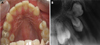
A 19-year-old male patient of Malay ethnicity was referred to our department for the treatment of a decayed upper right second premolar (Fig. 1A). Initial periapical radiograph confirmed endodontic involvement of the offending tooth. Additionally, it revealed the presence of two radiopacities in the maxillary canine-premolar region (Fig. 1B). The first radiopacity was cast due to the presence of a paramolar, whereas the second radiopacity gave an image of a transversely positioned "Y" shaped structure, consisting of supernumerary crowns and a short root and root canal, suggestive of a geminated supernumerary tooth. A pericoronal radiolucency of approximately 2 mm surrounding the geminated crown was found.
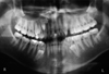
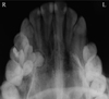
Panoramic radiograph revealed the presence of supernumerary teeth in both the maxilla and mandible (Fig. 2). To gain further insight into the buccolingual positioning of the supernumeraries, maxillary and cross sectional mandibular occlusal radiographs were recommended (Fig. 3).
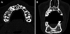
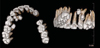
After obtaining informed consent from the patient, computed tomographic (CT) examination was performed with a 64-slice multidetector computed tomography (MDCT) scanner (Brilliance CT 64-channel, Philips Healthcare, Andover, MA, USA) using the dental software program DentaScan (GE Healthcare, Milwaukee, WI, USA). CT slices of 0.7 mm thickness were obtained (Fig. 4) along with the 3D reconstructed images (Fig. 5) which revealed the precise internal and external morphology of the supernumeraries. The 'Y' shaped structure consisted of a common root and root canal for three crowns: a medially directed molariform crown with a slight widening of the pericoronal space and two partially separated "premolar-like" crowns directed inferiorly, thereby confirming the diagnosis of geminated supernumerary tooth with trifid crown. Loss of the cortical plate of the palatal vault was also evident in the axial slices.
Owing to the possibility of causing interference in the restorative treatment plan, the erupted maxillary paramolar was extracted, whereas the other impacted supernumerary teeth, except for their slight pericoronal radiolucency, displayed no associated pathology or danger to the adjacent vital structures. Hence, a decision was made to keep the patient under periodic follow-up evaluation.
Discussion
Variation in the form and number of teeth is not an uncommon finding in clinical practice; however, their simultaneous presentation is a rare phenomenon. This report shows the occurrence of gemination in a supernumerary tooth, which to our knowledge is extremely rare. On reviewing the literature, we came across only two reports of geminated supernumerary teeth. Liu et al9 first described the occurrence of gemination in a supernumerary tooth in the mandibular premolar region in 2007 and proposed a new morphologic class "geminated-premolar-like" for the same. Yang10 reported a case of a geminated supernumerary tooth with two crowns and one root in the maxillary premolar region in 2012.
Our case was unique in that gemination of the supernumerary resulted in the formation of three crowns with a common root and root canal. After a thorough review of the literature and conducting a PubMed search using the keywords "gemination" or "geminated supernumerary" or "trifid tooth" or "triple tooth", we did not come across any report of a geminated supernumerary with three crowns. Interestingly, there was a similarity between this case and the one reported by Yang10 in terms of demographics of the patient, i.e. race (mongoloid) and gender (male). This could be a coincidental finding or might be an indication towards an increased prevalence of geminated supernumerary in this population, taking into account that supernumerary teeth tend to occur with high frequency in mongoloid males.11
As approximately 75% of supernumerary teeth are asymptomatic and remain impacted,12 they often present as an incidental finding on routine radiographic examination. Although conventional radiographs are usually able to provide sufficient details, they fail to provide definitive information concerning the 3-dimensional relationship of the supernumerary teeth and the surrounding structures for surgical planning.13 In our case, computed tomography (CT) was able to clearly reveal the intra-osseous location and morphology of the supernumerary teeth, as well as their proximity to the adjacent teeth, sinus, and cortical bone. Although MDCT was taken in this case, cone beam computed tomography (CBCT) would be a superior alternative in terms of radiation exposure.14
Surgical intervention for the removal of supernumerary teeth may be considered only if they pose a risk of cystic transformation and show a potential to cause local disturbances such as root resorption, rotation, and malformation of permanent teeth.15 In order to prevent future complications in cases of impacted supernumerary teeth, periodic follow-up evaluations are essential.
To conclude, geminated supernumerary teeth might often go undetected in routine dental practice, which can be attributed to their subtle presentation and the limitations of conventional radiography. Therefore, it seems worthwhile to draw more attention to gemination in supernumerary teeth and call for keen interest on the part of the dentist to recognise and report such cases with greater frequency.
 XML Download