

 PDF
PDF ePub
ePub Citation
 Print
Print


Introduction
The styloid process is a cylindrical, long cartilaginous bone located on the temporal bone in front of the stylomastoid foramen. Many nerves and vessels such as the carotid arteries are adjacent to the styloid process.1 The normal styloid process length is approximately 20-30 mm.2 The styloid process tapers toward its tip that lies in the pharyngeal wall lateral to the tonsillar fossa. Many important neurovascular structures lie near the tip of the styloid process. The internal carotid artery, internal jugular vein, and cranial nerves X, XI, and XII lie on its medial side.3 Keur et al4 suggested that a styloid process longer than 30 mm was considered to demonstrate styloid process elongation. The tip of the styloid process is continuous with the stylohyoid ligament, which extends to the lesser cornu of the hyoid bone. The stylomandibular ligament also attaches to the styloid process extending to the angle of the mandible. The protrusive movement of the mandible can be restricted by the stylomandibular ligament.5
Styloid process elongation is known as Eagle syndrome when it causes clinical symptoms such as neck and cervicofacial pain. It is assumed that these signs and symptoms originate from the compression of the styloid process on some neural and vascular structures. More uncommonly, symptoms such as dysphagia, tinnitus, and otalgia may occur in patients with Eagle syndrome. It may also cause stroke due to the compression of the carotid arteries.2
Thus, precise knowledge about the anatomy of both normal and abnormal styloids is important for clinicians, surgeons, and radiologists. Hence the present study evaluated and classified the radiographic appearance of styloid process calcification according to gender and site. Also, the clinical relationship between styloid process elongation and the limits of mandibular protrusion were investigated.
Materials and Methods
This study was conducted in the outpatient department from January 2010 to June 2011, during which 2,706 adults belonging to the Mathura region were investigated. The study was conducted according to the Human Ethics Guidelines approved by the institution, following which only those subjects were included for whom clinical examination and panoramic radiographs were prescribed as a part of a diagnostic work up.
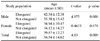
The subjects comprised 1,681 males and 1,025 females with a mean age of 34.54±14.62 years. The mean age of the male and female populations were 35.38±15.42 and 34.12±14.27 years, respectively. At the time of the study, all of the subjects were asymptomatic on styloid process palpation in the tonsillar fossa region.
As the stylomandibular ligament might restrict excessive protrusive movement of the mandible if the ligament were mineralized and elongated, we assumed that mandibular protrusive movement could be influenced. The mandibular protrusive movement, that is, the position of the mandible anterior to centric relation, was measured for all of the subjects by asking them to slide the mandible forward as far as possible. The distance was measured between the superior and inferior incisors in millimeters and the amount of horizontal overlap was added to it. The normal limit was ≥7 mm.5
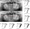
All the digital panoramic radiographs were taken by using a Planmeca ProMax X-ray unit (Planmeca Oy., Helsinki, Finland) under the standard exposure factor as recommended by the manufacturer. The type and calcification pattern of the right and left styloid process were analyzed independently. Any styloid process extending below an imaginary line connecting the anterior nasal spine and the mastoid process was considered elongated (Fig. 1).6
The styloid process elongation was recorded as unilateral or bilateral, and classified according to the system of Langlais et al (Figs. 1C and D).7 The study population was further divided into 4 groups as Group 1: not elongated; Group 2: elongated left side; Group 3: elongated right side, and Group 4 as bilateral elongation of the styloid process.
The relationships between the styloid process elongation on the one hand and gender, age, and mandibular movements on the other were investigated. The panoramic radiographs were examined by one investigator. The data were analyzed by using a Student's t-test and chi-squared tests. All analyses were executed using SPSS 15.0 (SPSS Inc., Chicago, IL, USA).
Results
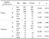
Out of a total of 2,706 panoramic radiographs, 1,295 (47.9%) were not elongated (Group 1), 141 (5.2%) showed left side (Group 2), 147 (5.4%) showed right side (Group 3), and 1,123 (41.5%) showed bilateral (Group 4) elongation of the styloid process (Fig. 2).
A calcified styloid was more prevalent in older subjects and males. The mean age was significantly higher in the patients with styloid process elongation (39.87±12.27 years) than the patients without styloid process elongation (34.54±14.62 years) (p=0.000, Table 1). Among the Mathura population in Group 2, 3, and 4, the most frequent type of calcification observed was elongated (Type I) and pattern was partially calcified (Pattern B) as shown in Figure 3 (Table 2). No statistically significant differences were proven between the styloid process types, calcification, and patterns among the subject gender and site prevalence (p>0.05), except for Group 4 where a statistically significant correlation was found in gender distribution for the left side and only for the pattern of calcification for the right side (Tables 2 and 3).
Further, the group of normal styloid process length bilaterally (Group 1, n=1,295) demonstrated mandibular protrusive movement to be 9.77±1.53 mm, whereas the elongated groups of Group 2 (n=141), 3 (n=147), and 4 (n=1,123) revealed limits of mandibular protrusive movement that were less extensive but still within normal range: 8.47±0.57, 8.42±0.55, and 7.56±1.11 mm, respectively. However, there was no statistically significant difference between styloid process elongation and mandibular protrusive movements (p>0.05, Table 4).
Discussion
An awareness of clinical and radiologic presentation of styloid process elongation is important to all health practitioners involved in the diagnosis and treatment of head and neck pain. Eagle syndrome, sometimes called styloid or stylohyoid syndrome, is defined as the symptomatic elongation of the styloid process or mineralization (ossification or calcification) of the stylohyoid ligament complex.8 It was first documented by Eagle, an otorhinolaryngologist, in 1937.3
Styloid process palpation in the tonsillar fossa, which is not normally palpable, is indicative of styloid process elongation. Palpation of the tip of the styloid process should exacerbate the symptoms associated with this syndrome. If highly suspicious for Eagle syndrome, it can be confirmed by radiographic imaging.5 There are many vessels, such as the carotid arteries, and nerves adjacent to the styloid process. The signs and symptoms of this syndrome originate from the anatomic relationship between the styloid process and its surrounding structures.9 The symptoms can be confused with some other disorders including a wide variety of facial neuralgias, and oral and temporomandibular diseases.10 Therefore, a detailed differential diagnosis for styloid process elongation should be performed.
Panoramic radiograph is a common modality for evaluating styloid process elongation. Skull lateral view is the best to show the length of the styloid process, but anteroposterior views are also needed to determine whether there is bilateral involvement and the presence of lateral deviation. Spiral CT with subsequent 3D reconstruction is the method of choice for exact determination of the localization of the styloid processes.11
Apart from the Eagle syndrome, other clinical manifestations that may result from calcified styloid process are glossopharyngeal neuralgia, carotidynia, pulsatile tinnitus, dysphonia, and globus pharyngeus. The pathophysiology behind the pain due to an elongated styloid process was found to be compression of the neural elements such as the glossopharyngeal nerve, lower branch of the trigeminal nerve, and/or the chorda tympani, by the elongated styloid process. The compression further leads to proliferation of granulation tissue that causes continuous pressure on surrounding structures and results in pain.12
Various theories have been proposed to explain the ossification of the stylohyoid and stylomandibular ligaments, namely the theory of reactive metaplasia, reactive hyperplasia, anatomic variance, ageing, developmental anomaly, and trauma leading to loss of elasticity in the ligament simulating tendinosis.13 The regional factors like dietary factors are also important for different patterns and types of styloid process elongation.14 The calcification of the styloid is now considered to be a part of heterotopic bone formation or ossification15 since, microstructurally, osteoid matrix is also present with the calcification.
Accurate determination using two-dimensional radiographic procedures can be difficult due to projection geometry considerations. Panoramic radiography may distort the dimensions of styloid process and magnification of the radiographic image may vary with the angulations of the process itself.6 Therefore, a simple measurement in millimeters is not a suitable criterion due to the radiologic factors involved. Taking this factor into consideration, to determine the length of styloid process, ruler measurement was not chosen.5 The elongation of the styloid processes in the radiographs was determined according to the method proposed by Ferrario et al.6
The present study was performed in the Mathura region of Northern India where the prevalence of elongated styloid process combining both unilateral and bilateral instances, was higher (52.1%) than those in previous studies on other Indian populations. A study was performed on the population of the Davangere region of Southern India16 where 173 panoramic radiographs were reviewed and the incidence of mineralization of the styloid process was found to be 24.8%. Also, More and Asrani studied 500 digital panoramic radiographs of a population in the Gujarat region of Western India, and they found elongated styloid process in 19.4% of its subjects.13 Both these investigators favored the theory of ageing for styloid process elongation.
However, in our study, the higher prevalence of elongated styloid could be attributed to the combination of factors including race, lifestyle, and dietary habits. The Mathura region mainly comprises a rural population where most people perform strenuous work like carrying heavy weight on their heads, which in turn promotes ossification in the ligament.17 They also chew hard foods like gutka and areca nut, which increases the masticatory load on the ligament and this leads to ossification of the ligament.18 In addition, racial predisposition may favor ossification of the ligament as the ossification center varies.19
We observed that the calcified styloid was more prevalent in older subjects, which was in accordance with the previous findings that with ageing there was progression in styloid process elongation.13 Additionally, we noted that the male population had a predilection for styloid process elongation. This result might be supported by the previous results of another study in an Indian population.13
Our study showed that for elongated styloid process, 79.5% of cases were bilateral and 20.5% of cases were unilateral, which was in agreement with the previous findings in which a bilateral elongation in 75% and a unilateral elongation in 25% of cases was reported.13 In the present study, the prevalence of Type I and partially calcified styloid process was >55%. These findings were similar to the study performed in the Gujarat population in India where the prevalence was >85%.13 The bilateral styloid process elongation could be attributed to the fact that strenuous exercise and chewing habits leads to clenching of the jaw muscles, which increases the weight loading of the styloid bilaterally and promotes ossification.
In our study, no statistically significant difference was observed between genders in the unilateral or bilateral elongation of the styloid process. This was in agreement with the reports of previous studies.6,20,21 Our hypothesis suggested that elongation of the styloid process had no effect on mandibular protrusive movements, which was similar to the findings of a study in a Turkish population.5 This could be attributed to the fact that all the patients included in the study were asymptomatic, which was similar to a previous study that found that despite a 84.8% prevalence of styloid process mineralization, none of the subjects were symptomatic.6 Langalis et al7 and Fini et al22 also reported a prevalence of subjective symptoms of only 1-5%.
Further, the ligament responsible for styloid process elongation might be the stylohyoid rather than the stylomandibular.6,23,24 It is not easy to determine the related ligament in cases where the elongated styloid process does not extend to the angle of the mandible. Correll et al25 and Zaki et al26 used the term "mineralization of the stylohyoid-stylomandibular ligament complex" for elongated styloid process on radiographs. We also recommend the term "mineralized stylohyoid-stylomandibular ligament complex" in view of the ambiguity of these structures when viewed on panoramic radiographs.
In conclusion, panoramic radiography is useful for detection of an elongated styloid process in patients with or without symptoms. Styloid process calcification in older adults is common with no correlation to gender. "Type I" with "partially mineralized" styloid process was observed more frequently in the population studied. Since the prevalence of an elongated styloid process was found to be higher in this study, awareness could be spread among people who are prone to styloid process elongation, especially rural populations and chronic gutka chewers. Further advanced imaging studies are required to identify the symptoms that correlate with an elongated styloid process as well as the type and pattern of calcification based on different lifestyles and dietary habits.


 XML Download