

 PDF
PDF ePub
ePub Citation
 Print
Print


Introduction
As a chronic infectious disease, dental caries is one of the most common problems encountered in clinical dentistry,1 and shows a very high incidence of over 80% of all adults in Korea according to the 2006 national survey.2 Early and accurate diagnosis of caries is essential for clinicians, who require exact knowledge of the depth of caries in order to determine the appropriate type of restoration and treatment planning.3,4 Among the various types of methods in the diagnosis of caries, probing, visual examination, intraoral film, and digital sensors are commonly used in routine clinical practice.5 Such diagnostic methods in the management of dental caries are used to determine the presence of caries and its extent, to monitor the course of caries progression, and to evaluate the effectiveness of treatment.6 However, a general observation from a review was that current methods tended to be more specific than sensitive (that is, their use results in relatively more false negative findings than false positive findings).7 Actually, several studies have shown that between 25% and 42% of caries lesions remain undetected by clinical examination performed without radiographic examination.8-10
When it comes to visual examinations with the adoption of the International Caries Detection and Assessment System(ICDAS II) clinical criteria in 2005, it has been attempted to correlate the clinical image of teeth to their histological status.11 However, the impact of these criteria on the diagnostic performance of visual examination is being examined on a limited number of studies, while the differences in caries location, lesion extent and experimental setup make extrapolation conduction difficult.12-15
In everyday clinical situation, if we exclude the recently introduced sophisticated methods for diagnosis of carious lesion such as fiberoptic transillumination (FOTI), electrical conductance (EC), laser fluorescence, and so on, the radiographic examination is the most frequently recommended method as a supplement to the clinical inspections.16,17 Regarding proximal non-cavitated caries, in spite of the variety of diagnostic modalities available, radiography is the most widely used method.11 Bitewing radiography has been available for more than 80 years,12,18 while in the more recent years digital radiographic modalities attempted to substitute conventional film radiography. The advantages of a digital system are the abilities to manipulate the image contrast and brightness and to magnify the images. However, extensive researches regarding conventional and various digital radiographic modalities failed to detect significant differences in their diagnostic performances, only to show comparable results.19-28
Despite its usefulness, the detection of caries lesions through conventional radiograph remains rather an elusive process.29 The limitations inherent in conventional radiography are mainly due to the 2D representation of caries lesions, which are 3D structures in reality, and this might lead to loss of valuable information.30,31 Small lesions remain undetected when the relative amount of mineral loss is low, resulting in low subject and image contrast. Moreover, the radiographic appearance of a lesion can change dramatically as a function of the chosen projection geometry. The replacement of film by digital detectors does not address these fundamental limitations.21,28,32,33
Dentistry has largely used the same method of 2D imaging since the first intraoral radiograph obtained in 1896. According to the review by Tyndall et al,34 only one or two significant advances in dental imaging have been made since then in the sense of imaging geometry. These advances include panoramic imaging and tomography, with the former being far more useful for dental applications, and the latter historically being limited primarily to temporomandibular joint and implant site imaging.34
Computed tomography (CT), which was invented by Hounsfield35 in 1973, is considered as a technical break-through. It is a well-known medical technique for the nondestructive examination of internal structures and its introduction to the dentistry has been revolutionary in the sense that it can provide true 3D imaging.29 Cone beam computed tomography (CBCT) is a new application of CT that generates 3D data at lower cost and absorbed doses than conventional "fan beam" CT found in the practice of medical field.36 Data from the craniofacial region are often collected at higher resolution in the axial plane than those from conventional CT systems.37 In addition, these systems do require relatively small amount of space and can easily fit into most dental clinics today. Although most of the CBCT usage has been confined to the applications for dental implant placement, orthodontics, surgery, and temporomandibular joint disease38-44 so far, several studies focusing on the diagnosis of dental caries have been reported.34
The aim of this article is to review the brief history of the usage of 3D concept in diagnosis of the dental caries and the current status of diagnosis using CBCT. This review builds on the findings of several recent articles related to caries diagnosis using CBCT, and seeks to outline the possible advanced applications that might be used in the future.
Materials and Methods
A PubMed search from 1965 up to February of 2011 was conducted to identify articles published in dental literature, and limited to human trials, using the search terms "caries", "diagnosis", "3 dimensional", "tomography", and "computed tomography". Manual searches of the bibliographies of all full text articles and related reviews selected from the electronic search were also performed.
Results
Early trials to realize the 3D image
An ideal diagnostic tool would enable the clinician to accurately assess the presence or absence of a lesion, to quantify its size and depth, and to determine its activity. Whereas the physics underlying the radiographic image formation process is well suited for imaging the dental structures, the sampling level of traditional intraoral imaging is not sufficient to fulfill these requirements. In order to acquire 3D information, the level of sampling needs to be increased.29
Earlier attempts have been made to improve the diagnosis of dentoalveolar conditions with 3D imaging using variations of tomosynthesis.34 This has been the underlying premise of tuned aperture computed tomography (TACT), which uses a limited number of basis projections to generate 2D slices at various depths.45 Although TACT provided some incremental benefit for periodontal and endodontic applications, improvements in caries detection and characterization were limited to simulated recurrent caries. In case of proximal lesions, a significant increase in detection rate could not be demonstrated according to several reports.46-52
Unfortunately, TACT has provided limited application in the practice of dentistry thus far, partly due to the advent of CBCT, ironically. The development of CBCT has been innovative because complete (360°) sampling is now possible without increasing the patient dose to unacceptable levels.29
A benchtop-based CT device using an intraoral detector as the image receptor, which was developed by van Daatselaar and coworkers,53 was another attempt used in caries diagnosis. It is referred as a local computed tomography (LCT), and has similar basic working principles with the commercial CBCT systems on the market, although it is not automated. They used a high resolution charge-coupled device (CCD) detector and rotating turntable with a fixed anode intraoral radiograph source. Both TACT and LCT generate a series of images that can be reconstructed into a series of cross-sectional images. Whereas TACT uses a fixed object and CCD sensor45 while moving the X-ray source, LCT uses a fixed CCD sensor and source and a rotating object.54
In their studies,53-56 they have shown the feasibility of LCT and the improved accuracy in caries detection compared with conventional radiography. The term, LCT is somewhat confusing with the local cone-beam computed tomography (LCBCT), which is frequently used in contrast to full volume CBCT. Recently, Kalathingal et al29 used LCT in their research and found no difference in the detection of carious lesions, however they did find that LCT was superior for assessment of caries depth.
Researches using clinical CBCT systems
Astounding speed of development and improvement of dental imaging technology makes the proper verification difficult. When it comes to application of clinical CBCT systems in dental caries research, the study of Akdeniz et al57 was the first English literature available via Pubmed search. They found that Accuitomo 3DX (Morita Co. Ltd, Tokyo, Japan) CBCT system was superior in caries depth assessment compared with conventional film radiography or storage phosphor (SP) images. There were many studies5,6,25,57-61 dealing with the caries researches using clinical CBCT system including the study by Akdeniz et al.57 Caries researches have been traditionally classified according to the location of caries lesion. Their studies dealt with only proximal lesions,5,6,57,59 occlusal lesions exclusively,60 and both proximal and occlusal lesions.25,58,61 Various CBCT systems with different settings have been used, although Accuitomo 3DX was the most frequently used one. They were compared with the conventional imaging modalities which included film radiography and digital intraoral radiography using CCD and photostimulable phosphor (PSP) plates. All the studies were in vitro studies, and they used premolar and molar teeth with various stages of caries lesions. Intraoral radiographs were taken using paralleling technique and rectangular collimation. For comparison of film radiography, the film was developed automatically. For the rating of the caries lesions, five-scale score was used when using histology examinations.
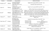
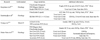
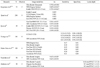
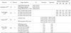
However, aforementioned studies have not been standardized in every aspect. For conventional imaging modalities, there were differences not only in systems but also in their X-ray taking environment such as voltage, current, exposure time, and focus to film (or receptor) distance. Regarding these differences, the authors might follow the manufacturer's instructions or clinical experiences. Storage method of teeth, the width of acrylic block mimicking the soft tissue, image software and comparison analysis were different among the studies. In addition, the status of caries lesion was diverse according to the studies. Several studies used the image of X-ray microcomputed tomography (MicroCT) as a gold standard instead of conventional histology. The number of observers was also different from study to study. Detailed information about imaging modalities and environments are described in Tables 1, 2, 3.
Methods of comparing analysis
Several studies5,6,59,61 used Az value for comparison which means area under the receiver operating characteristic (ROC) analysis. The ROC curve is a fundamental tool for diagnostic test evaluation. It allows to a complete sensitivity/specificity report. In a ROC curve the true positive rate (sensitivity) is plotted in function of the false positive rate (1-specificity) for different cut-off points of a parameter.62 Each point on the ROC curve represents a sensitivity/specificity pair corresponding to a particular decision threshold. The area under the ROC curve, Az, is a measure of how well a parameter can distinguish between two diagnostic groups (diseased/normal).63 Some aforementioned studies25,58 used sensitivity and specificity for comparing image modalities, however they did not present the ROC curve and Az values.
The study of Kamburoğlu K et al60 introduced the relative treatment effect (RTE) value as a statistical parameter according to the lesion depth. A 'treatment effect' is the average causal effect of a binary (0-1) variable on an outcome variable of scientific or policy interest. The term 'treatment effect' originates in a medical literature concerned with the causal effects of binary, yes-or-no 'treatments', such as an experimental drug or a new surgical procedure.64 However, the term is now used much more generally.
The study of Akdeniz et al57 is somewhat peculiar in that it compared the accuracy of determining the depth of proximal caries lesion, not just the accuracy of detecting the existence of lesion. Actually, this kind of studies, which deal with the defining the 3 dimensional region of the caries lesion, are now being actively performed by various research groups using X-ray microcomputed tomography instead of CBCT.65
Summary of comparisons
Tsuchida et al6 used the noncavitated proximal incipient lesions and found that no significant differences between CBCT and film images. This result might reflect difficulty of detecting incipient lesion.
Haiter-Neto et al25 compared NewTom 3G system with 3 fields of view as a full-volume CBCT with Accuitomo 3DX as a local CBCT (LCBCT). The intraoral radiography was also compared. The results showed that the NewTom 12-inches and 9-inches images had significantly lower sensitivities than the Accuitomo systems, whereas the NewTom 9-inches and 6-inches images had significantly lower specificities than the conventional radiography in detecting proximal lesions. The Accuitomo images were reported to be comparable with the conventional radiography. For occlusal caries detection, this LCBCT system presented a higher sensitivity than the other systems. In addition, it was determined to be equal to the intraoral systems, however the overall true score (true positives and negatives) was not higher for detection of dentinal lesions.
Young et al58 compared the efficacy of CBCT and conventional CCD image in detecting proximal and occlusal lesions. They used 3DX Accuitomo systems as a CBCT and found a significant difference in the average sensitivity score between CBCT and CCD regarding detection of proximal caries. They concluded that by using CBCT, it was able to improve the detection of proximal surface caries extending into the dentin, but not the occlusal caries.
Currently, there are two types of image detectors employed in CBCT equipments.59 One is the image intensifier with CCD, and the other is the flat panel detector including amorphous silicon flat panel and complementary metal oxide semiconductor panel. Qu et al59 evaluated the diagnostic accuracy of approximal lesions among the 5 clinical CBCT systems. The systems showed no statistical significant difference, and there was no significant difference between the types of the detectors of CBCT systems.
Kamburoğlu K et al60 assessed the diagnostic ability of intraoral digital CCD sensor images and CBCT images at different voxel resolutions in detection of occlusal caries. They found that the modalities of imaging performance were different in deep enamel, superficial dentin, and deep dentin. However, there was no difference in healthy and superficial enamel caries groups. They concluded that at all voxel sizes, CBCT images could be considered a tool for use in the diagnosis of occlusal caries.
The study by Şenel et al5 assessed the diagnostic ability of visual inspection, film, CCD sensor, PSP plate, and CBCT in the detection of proximal caries. The authors concluded that all the methods performed similarly in the detection of proximal caries.
Kayipmaz et al61 compared the effectiveness of conventional radiograph, PSP plate, and CBCT sytems in determination of occlusal and approximal caries. In determining occlusal caries, CBCT was statistically superior to the other two conventional methods. However, no significant difference was verified in determining approximal caries.
Discussion
Until now, only a small amount of research has been undertaken regarding caries diagnosis using CBCT. Although some studies maintained the superiority or the promise of using clinical CBCT system for diagnosis of dental caries, we could not find the consensus of the research up to now. There was a tendency among the studies to insist that the accuracy of CBCT systems was higher than the conventional methods in detecting occlusal caries and deep lesions into the dentin, however evidences are still insufficient. Sensitivity may increase, but mostly with simultaneous decrease of specificity. As mentioned above, the conditions of the experiments were different from study to study and not standardized. Thus, it is appropriate to say that whether the CBCT is superior to the conventional modalities in diagnosis of dental caries is under controversy at this stage. Furthermore, it goes without saying that the routine use of CBCT system instead of conventional radiography should not be accepted.
When considering a comparison of different modalities, it should be reminded that an increase in efficacy or lack of thereof does not always mean superiority or inferiority. We should consider cost, time, and effort. Above all, we cannot but consider the radiation dose despite the improvement of image.
The imaging of CBCT has fewer problems of geometric distortion than those of conventional methods in theory, thus the real 3D representation of caries lesion was available, which was impossible before. However, there are surely several limitations in clinical situations. It should also be reminded that all the studies mentioned were in vitro studies. The images were obtained under ideal geometry with no closed contact, cone cut, soft tissues, and projection distortions. In addition, the presence of any metal restoration in clinical situations might affect the quality of CBCT image. Further studies are compulsory not only to elucidate the accuracy of current systems, but also to verify and keep face with the future systems including in vivo studies.
 XML Download