

 PDF
PDF ePub
ePub Citation
 Print
Print


A face is an important aspect of the first impression a person casts upon another, thus a harmonious face is an integral component which may influence on the development of personality. Facial symmetry and overall harmony of the facial structures constitute an aesthetically pleasing appearance. The development of jaws is influenced by activities of the supporting musculature and the status of developing regional dentition. Any disruption in the equilibrium among these factors can conduct a disharmonized development. This may lead to facial asymmetry in significant proportions. A variety of causes manifest as facial asymmetry, among them chiefly are the dentofacial deformities. The main etiological factors that lead to dentofacial deformities can be divided into hereditary factors and environmental influences and known specific causes such as prenatal facial syndrome and congenital defects, or postnatal growth disturbance including effect of trauma.1
This report describes a patient with marked facial asymmetry due to a unique situation of developmental absence of few teeth and maxillary alveolar ridge on the left side. The effects of these disturbances on the facial musculature are also discussed. To the best of our knowledge, there has been no previous mention of such a case in English literature and this is the first case to be reported.
Case Report
A 17-year-old South Indian female was reported of a complaint of a pain in mandibular molar for three months. She had a very prominent facial asymmetry on the left side of her face. She had the asymmetry since the birth, however she was unaware of any progression of the asymmetry. She also reported of absence of both deciduous and permanent teeth in left posterior region of the upper jaw. There was no history of trauma or extractions in the region. Due to the absence of teeth, she never chewed on her left side. She also complained of a slight restriction in her mouth opening. Her family history was non-contributory. Her peri-natal history revealed a full term normal delivery and there was no history of any serious childhood illness.
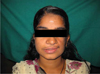
On general examination, she appeared to be in a good overall systemic health. Extraoral examination revealed a marked facial asymmetry with flattening of left middle third of her face. The left angle of her mouth was pulled upwards even at rest (Fig. 1). The lips were incompetent and mild drooping of left eye was seen. No abnormality was noted in her speaking, vision, and hearing. She exhibited a straight facial profile and a deficiency of muscle bulk on the left side of her cheek associated with hypertrichosis.
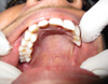
A slight reduction in mouth opening (approximately 25 mm inter-incisal opening) was noted. The mandible deflected to the right side with maximum mouth opening and maxillary midline shifted to the left side at rest position. Intraorally, hard tissue examination revealed clinical absence of all premolars and molars in the left maxilla. The corresponding alveolar ridge appeared rudimentary and underdeveloped (Fig. 2). The left deciduous maxillary canine was over-retained. The left mandibular first molar showed severe dental caries involving the pulp and exhibited tenderness on vertical percussion.
A panoramic radiograph of the jaws confirmed the absence of the left maxillary premolars and molars with severe underdevelopment of the left maxillary alveolar process. The lower jaw exhibited a full complete sixteen teeth (Fig. 3).
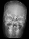
A posteroanterior projection of the skull also revealed the deficiency in the middle third of the face on the left side. No other abnormalities of the lower jaw or any other facial skeletal defects were noted (Fig. 4).
After confirming the absence of any other abnormalities, a diagnosis of segmental odontomaxillary hypoplasia was made. The patient was advised to have endodontic treatment of the dental caries in mandibular molar, followed by prosthetic rehabilitation of the maxillary missing teeth. The patient underwent the endodontic treatment, however chose not to have any intervention for the missing teeth or the facial symmetry as she had adapted to the situation.
Discussion
This case provided some unusual observations of regional congenital missing teeth with rudimentary type of alveolar process in the left maxilla. Also a prominent reduction of muscular bulk on the same side of the face causing facial asymmetry was notable although all other facial bones including mandible were normal. The alveolar process of the jaw bones develop in response to the presence of tooth buds. As the teeth develop and erupt, the alveolar processes develop and increase in height with bone deposition on the superior margin, which in turn grows in height and thickness of maxillary body. Therefore in our case, the absence of dental development seems to be the contributing factor in the failure of alveolar bone development in this region. The activity of a facial muscle primarily affects jaw growth in the following two ways; first, bone formation occurring at the point of muscle attachment depends on the muscle activity. Second, the musculatures are a part of the total orofacial soft tissue complex and they take a key role during jaws growth and maturation.1 In the present case, the patient had not been used the left side for the mastication because of left posterior maxillary teeth missing, and this disuse might have contributed to atrophy of left side facial musculature. Thereby, further bone growth of the upper jaw might be hindered.
Facial asymmetry may be a feature of various syndromes such as hemifacial microsomia, Crouzon's syndrome, facial clefting syndrome, and craniosynostosis syndrome.1 However, in this case there were no other abnormalities to suggest a syndromic manifestation. Except the left maxillary posterior alveolar ridge, all other facial bones including the mandible had normally developed.
Facial asymmetry due to a maxillary hypoplasia is a rare condition. Hemimaxillofacial dysplasia (HMD) and Segmental odontomaxillary dysplasia (SOD) are two other rare non-syndromic conditions causing facial asymmetry.2 In 1990, Danforth et al3 proposed the term "Segmental odontomaxillary dysplasia" for a more precise description of hemimaxillofacial dysplasia. The common features of these conditions were facial asymmetry, unilateral maxillary enlargement coupled with ipsilateral dental abnormalities such as congenitally missing molars and premolars and ipsilateral skin changes such as hypertrichosis or erythematous skin.4 They also demonstrated the posterior buccolingual expansion and dense sclerotic bone changes on the radiographs. Even though our patient also showed unilateral facial asymmetry, hypertrichosis and congenitally missing molars and premolars similar to HMD/SOD, buccolingual bone expansion and sclerotic bone changes were not observed. Instead, a rudimentary/hypoplastic posterior alveolar process on the left side was recognized clinically and radiographically. Our case might be designated as dysplasia, an abnormality of development. However, it was designated as hypoplasia due to her incomplete development of alveolar ridge, rather than abnormality.
In 1996, Packota et al5 analyzed the radiographic features of segmental odontomaxillary dysplasia by studying the radiographs of 12 cases. They found that the most common findings were missing premolar teeth, a smaller maxillary sinus, sclerosis and thickening of bony trabeculae, abnormal spacing of primary molars and vertical orientation of bony trabeculae. In 2000, Prusack et al2 reported a case of young child with facial asymmetry and reported a lack of right maxillary premolars, delayed eruption of right maxillary first permanent molar, abnormal shape of maxillary right primary molar roots, and decreased the size of right maxillary sinus. In contrast to our case, they also reported the enlargement of the corresponding alveolus associated with gingival hyperplasia. In 2003, Drake6 reported a case of 7-year-old boy with unusual maxillary asymmetry. His maxillary left premolars were missing. He also had superior canting of the left maxillary alveolus associated with insufficient vertical growth.
A thorough search of literature revealed a total of 31 cases of SOD/HMD reported until 2002,4 however to the best of our knowledge, segmental odontomaxillary hypoplasia has never been reported. The etiological factor for the present condition might be interrelated with the agenesis of left maxillary premolars and molars. This agenesis might cause the segmental odontomaxillary hypoplasia of left maxilla and disuse atrophy of corresponding facial musculature as we believe. Therefore, this report attempts to add another unusual cause of facial asymmetry, which is not yet been discussed in the literature.

 XML Download