Abstract
Purpose
Penile fracture is rare, but it is a urological emergency that always requires immediate attention. Moreover, penile fracture has been reported more frequently in recent years. It may have devastating physical, functional, and psychological consequences if not properly managed in time.
Materials and Methods
The objective of this study was to highlight the causes, clinical presentation, and outcomes of cases of penile fracture. This was a prospective observational study extending from November 2012 to November 2014. Each patient underwent a thorough clinical evaluation and received proper treatment.
Results
Twenty patients with penile fracture, aged 19 to 56 years (mean, 28 years) were evaluated in this study. Vaginal intercourse was the most common mechanism of injury. Most of the patients (95%) were diagnosed clinically with a proper history and clinical examination. Nineteen patients were treated surgically. The patients underwent six months of follow-up, and were evaluated with local examinations, questionnaires, and colour Doppler ultrasonography as necessary.
Conclusions
Although penile fracture is an under-reported urological emergency, its incidence is increasing. It is usually diagnosed based on a clinical examination, but ultrasonography can be very helpful in diagnosis. Especially in cases where treatment is delayed, surgery is preferable to conservative management, because it is associated with better outcomes and fewer long-term complications.
Although penile fracture has traditionally been considered a serious but rare urological emergency, its incidence has increased to the point that it can no longer be considered rare. Penile fracture is a misnomer; in fact, this condition is defined as a rupture of the tunica albuginea of the corpus cavernosum. The usual cause is abrupt bending of the erect penis by blunt trauma, which may occur during sexual intercourse, masturbation, rolling over on the bed, or falling onto the erect penis. The presentation of penile fracture may vary depending upon the time interval between occurrence and treatment and on the presence of associated injuries. Delay in presentation is mainly due to fear and embarrassment. The patient may recall hearing a cracking (pop-up) sound, followed by rapid detumescence of the erect penis and intense local pain. Hematoma, bruising, and deformity (known as 'eggplant deformity') of the penis then follow [12]. A palpable tunical defect and hematoma with a 'rolling sign' are usually considered pathognomonic features for this condition [3]. Associated urethral injuries may occur in a fair number of patients. The incidence of urethral injury is significantly higher in the USA and Europe (20%) than in Asia, the Middle East, and the Mediterranean region (3%), probably due to the different aetiology-intercourse trauma instead of self-inflicted injury [24567].
In the past, the diagnosis of this condition was usually clinical, based on high clinical suspicion and proper history taking. However, novel imaging techniques like ultrasonography (USG) [8], retrograde urethrography (RGU) [19], and the like help confirm the proper diagnosis when a diagnostic dilemma occurs [7]. As stated earlier, penile fracture may lead to devastating functional, physical, and psychological complications if not managed properly and in a timely manner [10]. The protocol for managing penile fracture has evolved from a conservative approach to the current standard of care involving immediate surgical exploration [14611]. The recommended procedure involves a degloving incision, evacuation of the haematoma, and repair of the rent of the tunica albuginea with absorbable or non-absorbable sutures [6]. Unsatisfactory penile curvature and erections, urethral strictures, and urethral cutaneous fistulae are among the complications that have been associated with the delayed treatment of penile fractures [412].
In our study, we analysed different aspects of penile fractures, including different modes of occurrence and presentation. Our study also addressed the management and outcomes of penile fracture, with special reference to the preservation of sexual function.
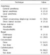
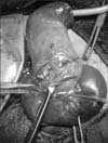
Institutional review board approval was taken for the study (IEC/230/12/02/2013). This was a prospective observational study extending from November 2012 to November 2014, including all patients admitted for blunt trauma to the erect penis. During this period, 20 cases of penile fracture were treated in our institute. Each patient underwent a thorough clinical evaluation and received proper treatment. Penile fracture was mainly diagnosed on clinical grounds, based on a proper history and clinical examination. USG was performed in 19 cases, and RGU was performed in one case. Both surgical and conservative treatment strategies were employed. Distal degloving was performed in 15 cases, and a direct lateral incision was performed in four cases (Fig. 1, 2).
Evacuation of the haematoma and repair of the tunical tear with absorbable sutures was carried out. Limited distal circumcision was performed in 12 cases. Perioperative catheterisation was performed in 16 cases, including the two cases involving urethral injuries. In 18 cases, six months of follow-up were completed., all patients were locally examined for penile deviation, fibrotic scarring, nodules, or other wound-related complication. In the third month after treatment, each patient's erectile function was evaluated. Patients' sexual function was evaluated using questionnaires and sexual function symptom scores, such as the International Index of Erectile Function-5 (IIEF-5) [13] or the Global Self-Assessment of Potency (GSAP) [14]. Both married and unmarried patients with a partner were evaluated with the IIEF-5, while unmarried patients without a partner were evaluated with the GASP. The IIEF-5 instrument classifies the severity of erectile dysfunction (ED) into five categories: severe (5~7), moderate (8~11), mild to moderate (12~16), mild (17~21), and none (22~25). The GSAP contains self-assessment questions about the severity of ED adapted from the Massachusetts Male Aging Study, with patients providing their own global self-rating for ED. ED severity was rated as none, mild, mild to moderate, moderate, or severe, depending on whether the patients were able to attain and maintain an erection adequate for satisfactory sexual intercourse always/almost always, usually, sometimes (approximately half of the time), infrequently (with only a minority of attempts at sexual intercourse being successful), or never, respectively. Colour Doppler studies were performed in patients with ED. Serial measurements of peak systolic velocity (PSV), end diastolic velocity (EDV), and resistive index (RI) were performed. Cavernous arterial insufficiency is likely when the PSV is <25 cm/s, as a PSV consistently >35 cm/sec defines normal cavernous arterial inflow. The vascular RI was defined as follows: RI=(PSV-EDV)/PSV. RI values >0.9 have been associated with normal penile vascular function, while RI values <0.75 are consistent with veno-occlusive dysfunction [15].
The patients were between 19 to 56 years old (mean±standard deviation, 33.35±11.99 years; median, 28 years). The time interval from injury to presentation was 6~156 hours (mean, 37.66 hours; median, 28 hours). The most common mechanism of injury was vaginal intercourse (50%). Masturbation (25%) and rolling over on an erect penis during sleep (25%) accounted for the rest of the cases (Table 1). When the penile fracture occurred, four of the patients were having sexual intercourse with the woman on top, three were watching an erotic film during masturbation, and two had ingested sildenafil tablets as a sexual stimulant. The injury occurred between 12 AM and 7 AM in 11 patients, six of whom were injured in the early morning hours, between 3 AM and 7 AM.
In a majority of the cases, the clinical presentation involved an audible popping sound (85%), followed by pain (50%), rapid detumescence (95%), and the development of swelling and discoloration (90%). Two patients experienced bleeding through the urethra. A typical 'eggplant deformity' was seen in 65% of the cases. A palpable gap in the penile shaft (the 'rolling sign') and a deviation of the penis to the opposite side of the fracture were seen in 55% and 65% of cases, respectively.
Diagnosis was possible on clinical grounds in 19 cases. One patient had a typical history, but the findings of a physical examination were not conclusive. USG was performed in 19 cases. A tunical tear was observed in 15 cases, and a tear of 2 to 3 mm was sufficient for diagnosis in the case in which a clinical diagnosis was not possible. RGU was performed in one case, in which the patient was suspected to have a urethral injury.
Surgical treatment was provided in 19 cases, while one case with a small tear was treated conservatively. A right corporal tear was observed in 12 cases, and 12 cases had a tear in the proximal third of the penis. Repair was performed using absorbable sutures in all cases (Table 2). Urethral injury was observed in two cases; in one case, the urethral injury was detected preoperatively by RGU, and in the other case it was detected during exploration through a distal degloving incision.
Two patients showed distal skin necrosis and were managed conservatively (Table 3). Follow-up was planned, involving a clinical evaluation during the third week and an evaluation of sexual function during the third month. At the first follow-up, all of the patients were evaluated, and two patients found to have a small nodule, which regressed spontaneously. At the second follow-up, 18 patients were evaluated, of whom 16 patients answered the IIEF-5 questionnaire (range of scores, 14~25; mean, 22; median, 22). Two patients complained of ED, with IIEF scores of 14 and 17, respectively (Fig. 3). On further evaluation, one of these patients was found to exhibit cavernosal insufficiency (PSV=25 cm/s) (Table 4).
The first documented report of penile fracture is credited to the Arab physician Abu al-Qasim al-Zahrawi in Cordoba, more than 1,000 years ago [11]. In the modern medical literature, the first case of penile fracture was described by Malis and Zur [16] in 1924.
The usual cause of penile fracture is abrupt bending of the erect penis by blunt trauma, which may occur during sexual intercourse, masturbation, rolling over in the bed, or during the practice known as 'taghaandan,' in which the erect penis is pushed down to achieve detumescence, resulting in a click [1]. The mechanism of injury depends on sociocultural characteristics, masturbation habits, and the specific sexual activities that an individual engages in.
The causes of penile fracture in our case series were similar to what has been reported in most other published series, with sexual intercourse being the most common cause.
Our literature review found that no data have been published regarding the time of occurrence of penile fractures. Most of the patients in our series were injured in the late night and early morning, which may reflect the circadian rhythm of testosterone secretion.
The diagnosis of penile fracture is often straightforward and can be reliably made through a proper history and physical examination, as in 95% of our cases. However, numerous recent studies have assessed the diagnostic role of various imaging modalities, such as USG [46817], cavernosography [618], RGU [19], and magnetic resonance imaging [619]. We found USG to be a very helpful tool in the diagnosis of penile fracture. USG was able to show a tunical fracture in 15 out of the 19 cases in which USG was performed in this study, and in one case, was able to show a 2~3 mm tear that confirmed the diagnosis despite an inconclusive clinical examination. In an article by Agarwal et al [11], USG was found to be sensitive in only 50% of cases. The results of USG are operator-dependent and USG requires specific expertise, which may explain the relatively poor results of USG in the previous study. RGU is highly sensitive, but is not essential for the diagnosis of urethral injury, since a suggestive history and proper surgical exposure with intraoperative retrograde instillation of methylene blue may be sufficient to diagnose urethral injury.
The protocol for managing penile fracture has evolved from a conservative approach to the current predominant approach that involves immediate surgical exploration [14611]. The surgical repair of penile fracture was first described by Fetter and Gartmen [20] in 1936 and became more popular in the 1980s [7], after several studies demonstrated that the rate of long-term complications was reduced from 30% to 4% in surgically treated patients [6]. Multiple contemporary publications have confirmed that suspected penile fractures should be promptly explored and surgically repaired. Muentener et al [10] compared surgical and conservative treatment strategies and reported success rates of 92% and 59%, respectively. Recently, Yapanoglu et al [21] and Gamal et al [22], in two similar studies, found that immediate surgical repair resulted in good outcomes and was superior to conservative treatment. In our series, surgical exploration was performed in 19 cases [16], while conservative management was employed in one case involving a small fracture with no signs of swelling or deviation. Hinev [23] has recommended conservative management when the cavernosal body is intact. Muentener et al [10] found that spontaneous healing without complications is probable for tears in the tunica albuginea without extensive haematoma or concomitant urethral injury, which may explain the outcome of our case. Agarwal et al [11] also reported a similar case in their case series. The conservative management of penile fracture has been associated with penile curvature in more than 10% of patients, abscess or debilitating plaques in 25% to 30% of patients, and significantly longer hospitalization times and recovery [24]. In sharp contrast to the abovementioned reports, the conservatively treated patient in our case series had a very good outcome. The proper selection of patients for conservative treatment may have led to the good outcome of conservative treatment in this case.
Penile fracture most commonly occurs on the right side and the ventrolateral aspect of the proximal third of the penis. The type and location of the incision is operator-dependent. Although small lateral incisions may be used for localized haematomas or palpable tunical defects [26], a distal circumcising (degloving) incision is appropriate in most cases, as advocated by Zargooshi [1], Miller and McAninch [9], and Mydlo [25]. In addition to being the most cosmetically favourable type of incision, distal degloving readily allows exposure to the entire tunica bilaterally, facilitating the diagnosis and repair of coexisting urethral and contralateral injuries. The decision to place a Foley catheter is operator-dependent. Some surgeons have reported routinely catheterizing their patients overnight, whereas others have advocated using a urethral catheter only when injuries are close to the urethra [56925]. The use of a catheter helps the intraoperative dissection without harming the urethra, facilitates the application of a pressure dressing, prevents postoperative wound contamination, and is unlikely to be harmful.
In uncircumcised patients, strong consideration should be given to performing limited circumcision at the conclusion of the repair procedure, because wide mobilization of the foreskin may place the distal prepuce at risk for ischemia [2]. We found distal skin necrosis in two out of three cases where a distal degloving incision was made but circumcision was not performed.
The differential diagnosis of penile fracture may include false fracture or rupture of the dorsal vein or the artery of the penis [262728]. An incidence of 4% to 10% false fractures has been reported [18], but we did not observe any such cases in our series.
The timing of surgery influences its long-term success. Patients undergoing repair within eight hours of injury have been found to have significantly better long-term results than patients who underwent surgery 36 or more hours after the fracture occurred [218]. In this study, the range of the time interval from injury to operation was 10 to 160 hours (mean±standard deviation, 43.27±38.06 hours; median, 31 hours). One patient underwent surgery 160 hours after trauma, and the only complication was a mild wound infection. The two patients who had ED in the follow-up were operated on 17 and 88 hours after injury. Thus, in our study, delays in surgery did not seem to have a particularly strong effect on the outcome.
Moreover, a lack of consensus exists regarding the need for postoperative suppression of penile erection with diazepam or oestrogen; this approach has been routinely used in some studies, but declared to be unnecessary in others [29]. The use of diazepam helps prevent early erections that might have harmful effects, and helps to allay the anxiety that may occur with such trauma. In our series, no definite protocol regarding the use of erectile suppressants was followed, and they were used according to the surgeon's preference. Supportive evidence in the literature was not available in this regard. However, pain during erection causes detumescence in and of itself, meaning that the use of such drugs is unnecessary.
The immediate postoperative outcomes also have varied in different case series. In our series, all patients were discharged on the third postoperative day, with the exception of four patients who developed complications. Two had mild skin infections and two had distal skin necrosis. All were managed conservatively and discharged between the fifth and tenth postoperative day. Different follow-up protocols and strategies have been reported in different published series. In this study, the first follow-up was in the third week after the operation, and all patients underwent clinical evaluation. Two had a small non-tender nodule over the injury site, and both nodules had resolved spontaneously by the next follow-up. Five patients had visible scars: four had direct lateral incisions and one had skin necrosis in the postoperative period. The next follow-up was at the third month, and only encompassed 18 patients. In this follow-up, postoperative sexual function was evaluated. Two patients had ED with low IIEF scores. On further evaluation with a Doppler study, one patient was found to have normal vascular flow and the other was found to exhibit cavernosal insufficiency. The most common causes of ED after penile fracture are corporeal veno-occlusive dysfunction, site-specific leaks, and cavernous artery insufficiency [30]. Zargooshi [5], in a personal surgical series incorporating 170 patients, reported that the surgical management of penile fractures resulted in erectile function comparable to that of a control population. A study performed by Nane et al [31], evaluating the long-term erectile status of patients in whom penile fracture was immediately repaired, noted ED in eight out of 36 patients after a mean follow-up period of 3.6±1.9 years. ED in the above patients was due to cavernosal and/or penile arterial insufficiency. Other reported complications include urethral stricture, urethra cavernosal fistulae [4]. A case of urethrocutaneous fistula following penile fracture has also been reported [12]. However, our prospective study did not contain any such complications.
Penile fracture is a urological emergency, should be managed promptly. Delay in presentation is mainly due to fear and embarrassment. Mechanism of injury depends on socio cultural characteristic, masturbation habits and indulgence in sexual activities. Diagnosis is usually clinical, but, USG is helpful. Surgery is the treatment of choice. However, conservative treatment may be given in properly selected patients. Early intervention gives better outcome, but, surgery should be offered in delayed presentation also to prevent long term sequelae.
The study limitation is, it has small numbers of patients with relatively short follow-up period. Studies with larger number of patients and a longer follow-up to detect long term sequelae of fracture penis are recommended.
Figures and Tables
Table 1
Patient characteristics and etiology (n=20)

Table 2
Surgical technique (n=19)

Table 3
Postoperative outcome (n=19)

Table 4
Follow-up after penile fracture repair (n=20)

References
1. Zargooshi J. Penile fracture in Kermanshah, Iran: report of 172 cases. J Urol. 2000; 164:364–366.

2. Morey AF, Dugi DD 3rd. Genital and lower urinary tract trauma. In : Wein AJ, Kavoussi LR, Partin AW, Novick AC, editors. Campbell-Walsh urology. 10th ed. Philadelphia: Elsevier-Saunders, Co.;2012. p. 2507. p. 2520.
5. Zargooshi J. Penile fracture in Kermanshah, Iran: the long-term results of surgical treatment. BJU Int. 2002; 89:890–894.

6. Jack GS, Garraway I, Reznichek R, Rajfer J. Current treatment options for penile fractures. Rev Urol. 2004; 6:114–120.
7. Derouiche A, Belhaj K, Hentati H, Hafsia G, Slama MR, Chebil M. Management of penile fractures complicated by urethral rupture. Int J Impot Res. 2008; 20:111–114.

8. Koga S, Saito Y, Arakaki Y, Nakamura N, Matsuoka M, Saita H, et al. Sonography in fracture of the penis. Br J Urol. 1993; 72:228–229.

9. Miller S, McAninch JW. Penile fracture and soft tissue injury. In : McAninch JW, editor. Traumatic and reconstructive urology. Philadelphia: W.B. Saunders;1996. p. 693–698.
10. Muentener M, Suter S, Hauri D, Sulser T. Long-term experience with surgical and conservative treatment of penile fracture. J Urol. 2004; 172:576–579.

11. Agarwal MM, Singh SK, Sharma DK, Ranjan P, Kumar S, Chandramohan V, et al. Fracture of the penis: a radiological or clinical diagnosis? A case series and literature review. Can J Urol. 2009; 16:4568–4575.
12. Mahapatra RK, Ray RP, Mishra S, Pal DK. Urethrocutaneous fistula following fracture penis. Urol Ann. 2014; 6:392–394.

13. Rosen RC, Riley A, Wagner G, Osterloh IH, Kirkpatrick J, Mishra A. The international index of erectile function (IIEF): a multidimensional scale for assessment of erectile dysfunction. Urology. 1997; 49:822–830.

14. Feldman HA, Goldstein I, Hatzichristou DG, Krane RJ, McKinlay JB. Impotence and its medical and psychosocial correlates: results of the Massachusetts Male Aging Study. J Urol. 1994; 151:54–61.

15. Naroda T, Yamanaka M, Matsushita K, Kimura K, Kawanishi Y, Numata A, et al. Evaluation of resistance index of the cavernous artery with colour Doppler ultrasonography for venogenic impotence. Int J Impot Res. 1994; 6:D62.
16. Malis J, Zur K. Der fractura penis. Arch Klin Chir. 1924; 129:651.
17. Hoekx L, Wyndaele JJ. Fracture of the penis: role of ultrasonography in localizing the cavernosal tear. Acta Urol Belg. 1998; 66:23–25.
18. Karadeniz T, Topsakal M, Ariman A, Erton H, Basak D. Penile fracture: differential diagnosis, management and outcome. Br J Urol. 1996; 77:279–281.

19. Abolyosr A, Moneim AE, Abdelatif AM, Abdalla MA, Imam HM. The management of penile fracture based on clinical and magnetic resonance imaging findings. BJU Int. 2005; 96:373–377.

20. Fetter TR, Gartmen E. Traumatic rupture of penis. Case report. Am J Surg. 1936; 32:371–372.
21. Yapanoglu T, Aksoy Y, Adanur S, Kabadayi B, Ozturk G, Ozbey I. Seventeen years' experience of penile fracture: conservative vs. surgical treatment. J Sex Med. 2009; 6:2058–2063.
22. Gamal WM, Osman MM, Hammady A, Aldahshoury MZ, Hussein MM, Saleem M. Penile fracture: long-term results of surgical and conservative management. J Trauma. 2011; 71:491–493.

26. Feki W, Derouiche A, Belhaj K, Ouni A, Ben Mouelhi S, Ben Slama MR, et al. False penile fracture: report of 16 cases. Int J Impot Res. 2007; 19:471–473.

27. Armenakas NA, Hochberg DA, Fracchia JA. Traumatic avulsion of the dorsal penile artery mimicking a penile fracture. J Urol. 2001; 166:619.

28. Bar-Yosef Y, Greenstein A, Beri A, Lidawi G, Matzkin H, Chen J. Dorsal vein injuries observed during penile exploration for suspected penile fracture. J Sex Med. 2007; 4:1142–1146.

29. el-Sherif AE, Dauleh M, Allowneh N, Vijayan P. Management of fracture of the penis in Qatar. Br J Urol. 1991; 68:622–625.


 PDF
PDF ePub
ePub Citation
 Print
Print


 XML Download