

 PDF
PDF ePub
ePub Citation
 Print
Print


Epidermal cysts are commonly encountered, slow-growing dermal or subcutaneous epithelial cysts that contain keratin and are lined by true epidermis [12]. Depending on the location of the lesion, these masses can be identified using ultrasonography (US), which is superbly suited to characterizing superficial lesions. They usually occur in the hair-bearing areas of the body, such as the scalp, face, neck, trunk, and back [123]. Epidermal cysts are a rare benign tumor of the testicle, and are usually discovered in the second and fourth decades of life [4]. We report an unusual testicular mass in an older patient, in whom the testis was found to have been completely replaced with an epidermal cyst.
CASE REPORT
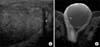
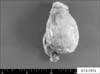
An 85-year-old man presented with a right scrotal mass noticed several months previously, and visited our hospital due to increased mild pain over the course of a week. The left testis and epididymis were intact. He had a history of controlled hypertension and no history of scrotal trauma or surgery. A physical examination of the testes found that the right testis was enlarged to approximately 10×6 cm, and a soft, non-tender, mobile, smooth-margined oval mass was palpable on the right scrotum. The transillumination test was negative. Scrotal US showed a mixed echoic mass mixed with a small, spindle-shaped echogenic reflector and a filiform anechoic area within the parenchyma of the right testis. The mixed echoic mass had totally replaced the right testis. Posterior echoic enhancement was also seen. On Doppler imaging, increased flow was not found in the mass (Fig. 1A). Magnetic resonance imaging showed a 9-cm, thin-walled, cystic oval component without internal enhancement, filling the entire right testis in the right scrotum (Fig. 1B). Radiologically, a testicular tumor, such as an epidermal cyst, was suspected. Enlarged lymph nodes were not observed. Serum alpha fetoprotein levels (3.94 mmol/mL) and beta-human chorionic gonadotropin levels (0.45 mIU/mL) were within normal limits. A right simple orchiectomy was performed for a presumed epidermal cyst. During the following 24 months, he underwent follow-up and reported no additional discomfort in the right scrotum. The pathologic report confirmed an epidermal cyst. The submitted specimen was large (9.0×6.2×4.5 cm, 166 g) and the outer surface was relatively smooth (Fig. 2). Grossly, the cut surface of the resected testis showed a unilocular cystic structure filled with light-grayish, keratinaceous debris replacing nearly the entire testis. The wall was evenly thick, measuring up to 0.3 cm across. The resected specimen was fixed in 10% formalin, paraffin-embedded, and stained with H&E. Entire sections were taken from the specimen.
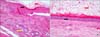
Microscopically, the cyst was observed to be lined by flattened squamous epithelium showing keratinization, and the cyst was filled with laminated materials resembling keratin. The surrounding testicular tissues and the adnexal tissues of the testis were noted to be normal, and no evidence was found of intratubular germ cell neoplasia or other dermal adnexal structures or teratomatous elements (Fig. 3).
DISCUSSION
Epidermal cysts commonly result from implantation of the epidermis into the dermis, as may occur in cases of trauma or during surgery. However, the exact cause is unknown. An epidermal cyst may be completely asymptomatic, or it may hurt when touched. It can release pus. It is very common for women to develop these cysts on the labia majora or minora. In contrast to pilar cysts, rare cases of malignancy can originate from epidermal cysts [5]. US is the standard for the imaging for most scrotal abnormalities, and is almost always the first imaging modality used to evaluate a testicular mass. Additionally, magnetic resonance imaging can be helpful in identifying signal intensities that conform to those of a simple cyst in order to confirm the diagnosis. However, epidermal cysts may occasionally be hyperintense to muscle on T1-weighted images due to proteinaceous cystic contents or intracystic lipid aggregates [6]. US usually shows a heterogeneous echoic density. Additional features include bright, strongly hyperechoic intralesional foci (echogenic reflectors), intralesional curvilinear or branchlike hypoechoic intralesional foci with a width of <0.3 cm (filiform anechoic areas), posterior acoustic enhancement, close apposition or mild bulging of the lesion into the overlying dermis without any intervening subcutaneous fat (dermal attachment), prominent focal bulging of the lesion protruding into the dermis, alternating hyperechoic and hypoechoic ring-like shadows (concentric ring pattern), and an echogenic center surrounded by a hypoechoic rim (target pattern) [7]. Pathologically, an epidermal cyst of the testis is classically defined as an intraparenchymal testicular cyst filled with keratinized material and lined by a complete or incomplete inner layer of squamous epithelium without teratomatous elements or cutaneous adnexal structures [8]. Generally, testicular-sparing surgery has been suggested [46]. The majority of patients are in their second to fourth decades of life. A review of the literature indicates that patients with epidermal cysts have been reported from three 77 years of age. The diameters of epidermal cysts have been found to range from 0.7 cm to 3 cm. However, the cyst in this case was 9 cm long [9]. Some reports have indicated that patient age does not correlate with the size of epidermal cysts [10]. In this case, the extremely large size of the cyst and the patient's advanced age suggest the possibility of tumor progression. Some researchers have also reported a progressively changing appearance of these masses. They followed their patients using US for up to six months. Over time, internal septae formed and the walls became thicker and irregular. This present case is unique in that it is the first report of an epidermal cyst in a patient more than 77 years of age, and the cyst was larger than any that have previously been reported. We believe that a testis totally replaced by an epidermal cyst should be resected due to the risk of progression and, although rare, the malignant potential of epidermal cysts.
In conclusion, in a rare presentation, an 85-year-old male patient was found to have an enlarged testis that had been totally replaced with an epidermal cyst. After surgical excision, no recurrence of an epidermal cyst of the testis was noted over the following 24 months.
 XML Download