

 PDF
PDF ePub
ePub Citation
 Print
Print


INTRODUCTION
Varicocele is detected in 2% to 11% of prepubertal boys [1,2,3] and 15% to 16% in postpubertal adolescent males [4]. Varicocele, the most common cause of secondary infertility in men, is a surgically correctable disease that causes deterioration in testicular function and semen parameters [5,6]. Varicocele has been found to be progressive with age [7]. Thus, the correction of varicocele at an early age may be recommended to prevent the deterioration of testicular function, including hypotrophy and infertility, considering the fact that prophylactic treatment is the best therapeutic method. Varicocele may become apparent peripubertally, and early corrective treatment may prevent damage to an individual's fertility status [8,9]. Moreover, increased testicular size after varicocelectomy has been reported in adolescents, but rarely in adults, although adults have significant increases in the total motile sperm count [8,10,11].
The treatments for varicocele include macroscopic inguinal or subinguinal varicocelectomy, angiographic embolization, microscopic inguinal or subinguinal varicocelectomy, and laparoscopic varicocelectomy [12,13]. It is not easy to accurately compare the outcomes of these methods due to innumerable and inconsistently controlled variables. Gubernacular veins were seen during varicocele surgery in 79% of the patients [14]. Murray et al [15] reported that gubernacular collaterals were presumed in 7% of varicocele recurrence.
Goldstein et al [16] have suggested that varicocelectomy with testicular delivery significantly reduces varicocele recurrence and postoperative hydrocele; however, few studies have compared varicocelectomy with and without testicular delivery [17]. Moreover, no controlled studies have been reported on varicocelectomy with testicular delivery in pediatric patients. Because varicocele in children is less common and is anatomically smaller, surgical methods in children have been developed only over the past two decades. This study examined the effects of magnification-assisted subinguinal varicocelectomy (MASV) performed with testicular delivery on recurrence and symptom resolution in children.
MATERIALS AND METHODS
1. Patient characteristics
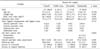
We retrospectively reviewed the medical records of 64 patients who underwent MSAV with testicular delivery at our institution between January 2010 and January 2014. Of these 64 patients, 23 were children 15 years or younger (mean age: 12.3±1.8 years; range: 8~15 years). Varicocele was observed on the left side and was found to be grade 3 in all 23 patients (Table 1).
Varicocele was diagnosed with a physical examination in the upright and supine positions by using the Valsalva maneuver. Varicoceles were classified as grade 1 (palpable only during the Valsalva maneuver), grade 2 (palpable without the Valsalva maneuver), and grade 3 (visible irrespective of palpation) [18]. Preoperative and postoperative testis sizes were measured with an ellipsoid Prader orchidometer (ASSI, Westbury, NY, USA) or scrotal color Doppler ultrasound (SCDU, HDI 5000; Philips, Bothell, WA, USA). If the difference in testicular volume was >2 mL or ≥15% on the affected side, it was defined as testicular hypotrophy. Unfortunately, we could not obtain semen data from our patients because they were too young to be assessed using a semen analysis. Operational indications for varicocele were scrotal discomfort or pain, testicular hypotrophy, and visible varicocele (grade 3) along with the patient's or parents' request or anxiety. We excluded patients with additional pathology of the scrotum or urinary tract (e.g., history of urinary tract infection, prostatitis, or epididymitis).
2. Varicocelectomy techniques
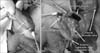
All procedures were performed under general anesthesia by a single surgeon, with the patients in a slightly head-down position [19]. Open MASV with testicular delivery was performed using standard techniques with the ligation of dilated veins. An incision was made below the external inguinal ring and the spermatic cord was identified (Fig. 1). The spermatic cord was mobilized to visualize the cremasteric muscle including the external spermatic veins (ESVs). ESVs were doubly ligated with 4-0 silk ties and divided. When the spermatic cord was reached, the cremasteric and internal spermatic fascia were opened in the longitudinal direction to preserve the external spermatic artery. Deferential vessels were first isolated and looped to preserve the deferential artery. Dilated internal spermatic veins (ISVs) were identified using 2.5× loupe magnification. Attempts were made to preserve the internal spermatic artery (ISA), and the vas deferens and deferential vessels, the cremasteric muscle, and the majority of the arteries were preserved as much as possible. The lymphatic sparing method was used for all the patients. The testicle was mobilized to allow the identification of gubernacular veins with intact gubernaculum. Small veins (less than 2 mm in diameter) were fulgurated, and larger veins were doubly ligated with 4-0 silk ties and divided. Before the procedures ended, the patients were changed to a slightly head-up position and the ipsilateral testicles were squeezed to check the remaining varicose vein. The cremasteric muscle was closed with interrupted absorbable sutures. The skin wounds were closed with subcuticular absorbable sutures and derma bond was applied. The mean operation time for MASV with testicular delivery was 73.5±17.5 minutes.
All patients were discharged on the following day of the surgery and were examined 1 week later to evaluate the wound. All patients were evaluated at 3, 6, and 12 months postoperatively, and every 12 months thereafter. The evaluations included a testicular examination of the scrotum with or without SCDU. Surgical success was defined as the absence of varicocele, and recurrence was defined as the presence of varicocele (≥grade 1) on the clinical examination after the surgery. All patients were followed-up for a minimum of 3 months; the mean follow-up time was 10.8±6.6 months.
3. Statistical analyses
The reason for surgery (discomfort, palpable mass and hypotrophy), ISA preservation, operation times (from incision to closure), success rates and symptom resolution rates were analyzed using one sample t-tests, Mann-Whitney U test, and analysis of variance (ANOVA), as appropriate. All statistical analyses were performed using SPSS version 12.0 (SPSS Inc., Chicago, IL, USA). All statistical analyses were two-sided, with p<0.05 considered statistically significant.
RESULTS
The surgical success rate of varicocele was 100%. The overall symptom resolution rate was 91.3% (Table 1). Varicocelectomy was decided upon due to scrotal hypotrophy (n=14, 60.9%), the existence of a mass (n=6, 26.1%, including 1 recurrent case), and discomfort (n=3, 13.0%). The scrotal mass and discomfort disappeared, but the testicular catch-up growth did not occur in 2 among 14 patients with scrotal hypotrophy. Fourteen of the 23 patients were diagnosed with ipsilateral testicular hypotrophy before treatment, although we were only able to compare the testicular size before and after successful MASV with testicular delivery in 11 patients. The left testis volume increased from 6.5±4.3 mL to 10.6±7.5 mL (p=0.003).
ISA preservation was successful in 8 patients (34.8%). There were no significant inter-group differences in terms of the surgical success rate, symptom resolution, and catch-up growth between the ISA preservation group and the ligation group.
Postoperatively, all patients experienced scrotal inflammation that lasted for 4 to 21 days, but the condition resolved over time. None of the patients revealed testicular atrophy or hydrocele.
DISCUSSION
Macroscopic inguinal or subinguinal varicocelectomy performed without optical magnification may miss smaller ISVs that may later dilate and cause varicocele recurrence [12]. Many urologists believe that the collateral reflux of spermatic veins may be a factor causing recurrence. Coolsaet [20] concluded that varicoceles was caused by reflux into the ISV (67% of the cases), the extrafunicular veins (20% of the cases) or both types of veins (14%). Microsurgical inguinal varicocelectomy with testicular delivery provides a direct visual access to all avenues of testicular venous drainage and is reported to result in a significant decrease in varicocele recurrence [14,16]. Only 1 available comparative trial revealed no statistical difference between surgery with testicular delivery and surgery without testicular delivery [17]. However, thus far, there have been no controlled studies on varicocelectomy with testicular delivery in pediatric patients. In the current preliminary study including pediatric patients, testicular delivery was also useful to easily ligate ESVs and gubernacular veins and was safe and without any complications. Therefore, additional comparative studies with larger samples are warranted to reveal the definite results.
Varicocelectomy using microscope magnification allows the identification of the testicular artery, lymphatics, and small venous channels, which aids in the preservation of the arterial and lymphatic vessels, and allows a complete ligation of the spermatic veins, which in turn minimizes the risk of postoperative complications. These methods significantly decrease the incidence of hydrocele formation, testicular artery injury and varicocele recurrence [21]. Park et al [22], who compared the data of adult and pediatric patients, concluded that there was no difference in the surgical difficulty and the microscopic subinguinal varicocelectomy truncated operative time in children. They stated that the omission of testicular delivery may be possible in prepubertal boys because gubernacular vein enlargement is not present in this population. However, we found that gubernacular veins ≥2 mm in diameter were to present in 17.4% (4/23) of the cases and smaller gubernacular veins were fulgurated in 3 cases. Murray et al [15] also found presumed gubernacular collaterals in 7% of varicocele recurrence. Despite the potential problems of scrotal hematoma and longer operative time following testicular delivery, there were no complications after MASV with testicular delivery in this study.
Microscopic equipment is very expensive, and a microscopic procedure is very delicate and microsurgical subinguinal varicocelectomy has a long learning curve. Loupe magnification is considered possible option for microscopic equipment for subinguinal varicocelectomy with testicular delivery. Loupe-assisted subinguinal varicocelectomy with testicular delivery is useful for collateral veins and for isolating ISVs from the remaining structure. However, we should remember that MASV using loupe plays a limited role in the isolation of ISA from the spermatic cord as compared to microscopic subinguinal varicocelectomy. In the current preliminary study, ISA preservation was successful in 8 patients (34.8%). This rate of ISA preservation was lower than 100% of subinguinal varicocelectomy using a microscope or 54% of laparoscopic varicocelectomy [22,23]. The total operation time in the current study was 73.5±17.5 minutes. This is comparable with the result (78±18 minutes, n=18) of the microsurgical subinguinal varicocelectomy in children and lower than 94±18 minutes in adults [22].
Varicocelectomy has been shown to be effective in the control of pain or discomfort [24,25]. One study found that 73% of the subjects had complete or significant resolution of discomfort and 10% had partial resolution after varicocelectomy, suggesting that surgical treatment is effective for painful varicocele [26]. In a Korean study, 78.6% of men experienced complete relief from discomfort and 9.7% had partial relief whereas 11.6% had persistent or worsened pain or discomfort [27]. These findings suggested that the postoperative degree of pain relief was affected by the preoperative quality of pain. We already reported that the complete resolution of testicular discomfort was seen in 76.7% of the patients, including 55.6% of the adolescents and 82.4% of the adults, after varicocele repair [28].
In the current study, catch-up growth occurred in 85.7% of the patients after MASV with testicular delivery in those treated with a 34.8% ISA preservation rate. Zampieri et al [29] reported that 80% of the patients demonstrated testicular catch-up growth within 18 months after surgical intervention, and only 32% of the patients demonstrated complete and real testicular volume catch-up. The high catch-up growth rate in this study compared with previously published studies may be due to the surgeries being performed at a relatively young age, possible edema effects in shorter follow-ups and the small number of children who were included in this retrospective analysis. Therefore, large-scale prospective studies with long-term follow-ups are necessary to estimate the real catch-up growth rate.
 XML Download