

 PDF
PDF ePub
ePub Citation
 Print
Print


The common characteristics of crossed testicular ectopia (CTE) are ipsilateral cryptorchidism or ectopic gonads and contralateral hydrocele with the absence of a testis [1-7]. In addition, hypospadias, pseudohermaphroditism, and scrotal abnormalities can also occur together [1,6]. Even though about 100 cases have been reported in the world literature, infantile CTE cases initially presenting as an incarcerated inguinal hernia are very rare.
CASE REPORT
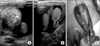
A healthy 10-month-old boy was admitted to the emergency room with a painful swelling in the left inguinal region. Physical examination showed a tender 2-cm lump in the left inguinal canal. Inguino-scrotal ultrasonography in this patient showed a left inguinal hernia with two gonads in the left scrotal sac and an impalpable right non-scrotal testis (Fig. 1A, 1B). In addition, we could not find evidence of persistent Müllerian duct syndrome, which is characterized by the presence of a uterus and fallopian tubes in abdominal ultrasonography. There was no evidence of developmental malformations of the external genitalia, such as hypospadias, bifid scrotum, or micropenis. After urgent manual reduction for an incarcerated inguinal hernia, left inguinal exploration was undertaken. A thin-walled inguinal hernial sac was found, containing two normal-sized and normal-shaped testes that were attached to one gubernaculum from the left inguinal ring (Fig. 1C). Each had its own blood supply and had separate vasa deferentia. After left inguinal herniorrhaphy, the gubernaculum was divided to strengthen each of the spermatic cord without creating undue tension on either side. The left testis was placed into the left subdartos space and the ectopic right testis was fixed transseptally into the right subdartos space.
DISCUSSION
This uncommon anomaly can occur together with persistent Müllerian duct syndrome or the presence of male pseudohermaphroditism, and an abnormal karyotype [1,6]. CTE has been classified into three types, based upon the presence of associated developmental abnormalities [1,2]. Type I, the most common, is associated with an indirect inguinal hernia. Type II is associated with the presence of persistent Müllerian duct structures, such as a rudimentary uterus or fallopian tubes. Type III is associated with other genitourinary abnormalities, such as hypospadias or scrotal abnormalities [1]. In this case, we could not find any ambiguity in genital phenotypes or any trace of a uterus and fallopian tubes in the pelvic cavity by ultrasonography. Furthermore, the architecture of both of the kidneys and the spleen was normal. On the basis of these findings, our case can be classified as type I.
CTE cases have rarely been reported in the English-language literature worldwide [1-7]. Our case is particularly notable because of the unusual presentation of CTE as an incarcerated inguinal hernia. To our knowledge, the present case represents the third case of CTE diagnosed in an infant with an incarcerated inguinal hernia in the world (Table 1) [3,4].
In most reported cases, the correct diagnosis of CTE is not appropriately made preoperatively because of its rarity [5]. Furthermore, because incarcerated inguinal hernia like this case should usually be immediately managed, a complete evaluation is usually not possible [2-4,6]. For this reason, the correct diagnosis is not easily made preoperatively. In our case, ultrasonography was very helpful for the diagnosis of CTE and for deciding upon the proper management [7].
Surgical procedures for contralateral orchiopexy have been described; contralateral orchiopexy can be performed through the scrotal septum (transseptal orchiopexy) [3,4] or through a suprapubic subcutaneous tunnel (through their respective superficial inguinal rings) [5].
Because the incidence of testicular cancer generally increases in fixed testes, careful follow-ups are imperative [8].

 XML Download