Abstract
A 26-year-old man complained of a vague low abdominal discomfort for the previous 2 months. Radiologic evaluations demonstrated that there was tubular structure connected with the right side wall of the bladder, suggesting Meckel's diverticulum with fistula formation to the bladder as well as a mass-like bladder wall thickening. With an impression of Meckel's diverticulum with fistula with the bladder, laparoscopic surgery was performed to confirm a diagnosis and to manage the Meckel's diverticulum with fistula with the bladder. The distal tip of the appendix was firmly attached to the right side of the bladder. The final diagnosis was corrected by laparoscopy followed by laparoscopic appendectomy and fistula repair. Vesico-appendiceal fistula is an uncommon type of vesico-enteral fistula and a rare complication of unrecognized appendicitis. Additionally, this report showed the significant value of laparoscopy as a diagnostic and therapeutic tool to this entity.
Vesico-appendiceal fistula is rare, with most cases related to benign or malignant disease. It is very difficult to diagnose preoperatively because the symptoms are vague and diagnostic tools cannot readily identify the disease. To date, there have been only 4 cases treated with laparoscopy. We report on a patient with vesico-appendiceal fistula treated by laparoscopy.
A 26-year-old man who appeared healthy was transferred to our facility for persistent intermittent low abdominal and perineal pain for the previous 2 months. The patient had also experienced urinary symptoms including frequency, urgency, gross hematuria, and dysuria for a month and had no history of any medical or surgical procedures prior to the visit. He was treated with antibiotics for 2 months under a diagnosis of cystitis and Meckel's diverticulum or Crohn's disease at local clinics; however, there were no pathologic findings on colonoscopy and no complaint about bowel symptoms. His family history was not notable for Crohn's disease.
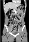
He was 177 cm in height and 72 kg in weight with no acute distress. On physical examination, there were no abnormal findings. There was pyuria and microscopic hematuria on urine analysis; however, the other laboratory findings were within normal range. Cystoscopy showed diffuse erythematous mucosal thickening on the right side wall of the bladder dome (Fig. 1). However, there was no stool debridement no any fistulous opening in the bladder. A computed tomography (CT) scan showed that there was a tubular structure connected with the right side wall of the bladder, suggesting Meckel's diverticulum with fistula formation to the bladder as well as a mass-like bladder wall thickening (Fig. 2).
With an impression of Meckel's diverticulum with fistula with the bladder, a laparoscopic operation was performed through 3 abdominal ports (one 10 mm port placed at 10 mm above the umbilicus [camera] (KARL STORZ GmbH & Co. KG, Mittelstr, Tuttlingen, Germany), one 12 mm port for the right pararectal trocar, and one 5 mm port placed between the left anterior iliac spine and the umbilicus). The distal tip of the long appendix, which had a normal shape on its body and base was found to be densely adhered to the right side wall of the bladder (Fig. 3). Laparoscopic appendiceal ligation was performed with 10 mm Hem-O-Lok clips (Teleflex Medical, Research Triangle Park, NC, USA) and 2-0 Vicryl (Ethicon Inc., Somerville, NJ, USA). After removing the proximal appendix, dissection of the bladder around the tip of the appendix was performed. There was a dense fibrotic change around the tip of the appendix. Partial cystectomy was performed and laparoscopic two-layered bladder repair was done with 3-0 Vicryl (Ethicon). The total surgical time was 75 minutes and the estimated intraoperative blood loss was minimal.
On the 7th postoperative day, cystography was performed and no urinary leakage around the bladder was demonstrated. The urinary symptoms including gross hematuria, dysuria, and frequency improved and abdominal discomfort symptoms also subsided. The other laboratory findings were within normal range. The surgical specimen showed a 9.2×1.2 cm appendix attached to a 4.3×3.2 cm bladder with dense fibrotic change without any malignancy.
At 1 month postoperatively, he had no symptoms including frequency, urgency, sense of residual urine, or intermittent gross hematuria. Nor were there any abnormal findings by CT which was performed 1 year postoperatively.
Vesico-enteric fistula is a rare complication of appendicitis and malignancy. The common causes of vesico-enteric fistula include colonic diverticulitis (51%), colorectal cancer (16%), Crohn's disease (12%), and bladder cancer (5%).1 Additionally, vesico-appendiceal fistula is extremely rare, constituting less than 5% of all vesico-enteric fistulas.2 It occurs most often in males between the ages of 10 and 40.3 The higher incidence of this disease in males originates from the uterine interposition between the bladder and appendix in females.
The most common symptoms included recurrent urinary tract infection and lower abdominal discomfort. The most common organisms cultured in urine are Escherichia coli and Klebsiella.3 The patient also demonstrated irritative bladder symptoms including frequency, dysuria, and intermittent lower abdominal discomfort. Occasionally, a patient complains of pneumaturia and fecaluria. However, we could not find pneumaturia and fecaluria in this case. It has been thought that the complete obstruction of the fistula is caused by fecalith. We found a fecalith in the appendiceal lumen intraoperatively.
A vesico-appendiceal fistula causes misreading acute appendicitis.4 However, early diagnosis is difficult, because the symptoms are vague and useful diagnostic tools have not yet been identified. It has been reported that it usually has taken at least 1 year from the onset symptoms to diagnosis.5 Many diagnostic tools, such as intravenous pyelography (IVP), cystoscopy, cystography, colonoscopy, and CT scans, have been used for detecting a vesico-appendiceal fistula.3 Cystoscopy is sometimes useful in patients showing a fistulous opening in the bladder wall. We found an edematous bladder wall without a fistulous opening in this case. Cystography is also a useful diagnostic tool in patients who demonstrate a fistula tract between the bladder and appendix. In our case, colonoscopy was useful to rule out intestinal pathology. To date, a CT scan has been recognized as the most accurate diagnostic test, whereas plain films and IVP are not helpful. Some authors have demonstrated that the thin interval of the CT scan section was helpful in identifying vesico-enteric fistula. We also found an abnormal fistula between the bladder and intestinal tract; however, the CT scan did not identify the origin of the fistula exactly, not distinguishing between Meckel's diverticulum and the appendix.
The treatment of vesico-appendiceal fistula consists of appendectomy and partial cystectomy. We removed large portions of the bladder dome and anterior wall because the inflammatory change was so extensive. Some authors have reported repair of the bladder wall with an Endo-GIA (Ethicon Endo-Surgery Inc., Blue Ash, OH, USA) stapler.3 Because this may cause infection and stone formation, we performed laparoscopic appendectomy and laparoscopic partial cystectomy with a double-layered bladder wall repair using 3-0 Vicryl.
Although more than a hundred vesico-appendiceal fistulas have been reported, laparoscopic management has been reported in only 4 cases up to the present according to our literature review.3,6 This case showed the value of laparoscopy as diagnostic and treatment modality in vesico-appendiceal fistula.
Figures and Tables
 | Fig. 1Cystoscopy showed bullous erythematous changes of the dome of the urinary bladder. However, there was no definite fistulous tract opening or stool debridement in the bladder. |
ACKNOWLEDGEMENTS
This work was supported by a clinical research grant from Pusan National University Hospital 2012.
References
1. Carson CC, Malek RS, Remine WH. Urologic aspects of vesicoenteric fistulas. J Urol. 1978. 119:744–746.

3. Chung CW, Kim KA, Chung JS, Park DS, Hong JY, Hong YK. Laparoscopic treatment of appendicovesical fistula. Yonsei Med J. 2010. 51:463–465.

4. Ikeda I, Miura T, Kondo I. Case of vesico-appendiceal fistula secondary to mucinous adenocarcinoma of the appendix. J Urol. 1995. 153:1220–1221.

5. Bigler ME, Wofford JE, Pratt SM, Stone WJ. Serendipitous diagnosis of appendicovesical fistula by bone scan: a case report. J Urol. 1989. 142:815–816.

6. Afifi AY, Fusia TJ, Feucht K, Paluzzi MW. Laparoscopic treatment of appendicovesical fistula: a case report. Surg Laparosc Endosc. 1994. 4:320–324.


 PDF
PDF ePub
ePub Citation
 Print
Print


 XML Download