

 PDF
PDF ePub
ePub Citation
 Print
Print


Inverted papilloma of the urinary tract is a rare benign lesion. More than 100 cases have been reported and most frequently have been found to occur in the bladder.1 To our knowledge, only 1 inverted papilloma of the prostatic urethra has been reported in Korea.2 Notably, we report the first case of inverted papilloma of the prostatic urethra arising in a juvenile in the literature.
CASE REPORT
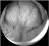
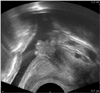
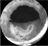
A 17-year-old man was referred for evaluation of painless, gross hematuria and voiding difficulty. The patient had suffered from voiding symptoms for 2 months. The patient did not have any history of trauma or infection. He had a no family history or personal history of urinary tract diseases. The physical examination was unremarkable. Urinalysis showed greater than 30 erythrocytes, 0 to 2 leukocytes/HPF, and a pH of 6.0. The urine culture was negative. The blood urea nitrogen and creatinine was 15 mg/dl (normal range 8 to 18) and 0.6 mg/dl (normal range 0.5 to 1.5). Cystoscopy revealed a solitary, papillary tumor on the prostatic urethra protruding to the bladder neck (Fig. 1). It was located on the prostatic urethra proximal to the verumontanum. Transrectal sonography demonstrated a 1.4 cm papillary lesion on the medial aspect of the prostatic urethra protruding to the bladder neck (Fig. 2). The inside of the bladder was unremarkable. Bladder washing cytologic specimens were negative for malignancy. Transurethral resection was then performed (Fig. 3) and a histological examination showed an inverted papilloma of the prostatic urethra (Fig. 4). At 3 months of follow-up, he was asymptomatic.
DISCUSSION
Urinary tract inverted papilloma is an uncommon urothelial neoplasm that demonstrates an inverted growth pattern, usually composed of anastomosing islands and trabeculae of histologically and cytologically normal urothelial cells invaginating from the surface urothelium into the subjacent lamina propria but not into the muscularis propria. The term "inverted papilloma" was initially introduced in 1963 by Potts and Hirst3 to describe this architecturally distinctive urothelial neoplasm.
Urothelial inverted papilloma is usually found in men in their sixth or seventh decade of life.4 However, in our patient, inverted papilloma was found in the second decade of his life. Sung et al5 reports that male predominance (male-to-female ratio, 7.3 : 1) and a mean age of 60 years were observed in their patients, though a wide age range (from 26 to 85 years) was noted. In addition, the majority of inverted papillomas developed in the urinary bladder (89%, 67 of 75 cases), particularly on the trigone (32%, 24 cases) and bladder neck (21%, 16 cases). Patients complained of hematuria (51%), dysuria (35%), or irritable symptoms (16%) and 24% had more than one symptom, in keeping with previous studies.4,6 Also, our patient complained of hematuria and voiding difficulty.
The inverted papilloma has no specific radiologic characteristics. Diagnosis requires direct visualization and biopsy. Cystoscopy can be used for both direct visualization of the lesion and entire local resection of a lesion.7 In addition, transurethral resection is regarded as the standard treatment for inverted papilloma in the lower urinary tract.7 We also performed transurethral resection to remove the prostatic mass.
Inverted papilloma of the prostatic urethra arising in a juvenile has never been reported. Furthermore, this case was of interest in that inverted papilloma of the prostatic urethra could be a factor in hematuria and voiding difficulty in juveniles. Here, we reported a rare case of inverted papilloma of the prostatic urethra, which was removed successfully.

 XML Download