

 PDF
PDF ePub
ePub Citation
 Print
Print


Abstract
Congenital thoracic kidney is a rare congenital malformation, caused by renal malpositioning during embryogenesis or congenital diaphregmatic hernia with herniation of kidney. Prenatal diagnosis of congenital thoracic kidney has been only rarely reported, underlying congenital diaphragmatic hernia should always be suspected in cases of congenital thoracic kidney. We present a case in which the prenatal diagnosis of an ectopic intrathoracic kidney was made on routine anatomical survey at 29 weeks' gestation.
Figures and Tables
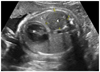
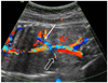
Fig. 1
Transverse sonograms of the fetal thorax on prenatal sonographic examination at 29 weeks demonstrate the ectopic kidney (K) in the left hemithorax, compressed left lung (L), mild right shift of the heart (H).
References
1. Sözübir S, Demir H, Ekingen G, Güvenç BH. Ectopic thoracic kidney in a child with congenital diaphragmatic hernia. Eur J Pediatr Surg. 2005. 15:206–209.
2. Panda B, Rosenberg V, Cornfeld D, Stiller R. Prenatal diagnosis of ectopic intrathoracic kidney in a fetus with a left diaphragmatic hernia. J Clin Ultrasound. 2009. 37:47–49.
3. Athanasiadis AP, Zafrakas M, Arnaoutoglou C, Karavida A, Papasozomenou P, Tarlatzis BC. Prenatal diagnosis of thoracic kidney in the 2nd trimester with delayed manifestation of associated diaphragmatic hernia. J Clin Ultrasound. 2011. 39:221–224.
4. Donat SM, Donat PE. Intrathoracic kidney: a case report with a review of the world literature. J Urol. 1988. 140:131–133.
5. Liddell RM, Rosenbaum DM, Blumhagen JD. Delayed radiologic appearance of bilateral thoracic ectopic kidneys. AJR Am J Roentgenol. 1989. 152:120–122.
6. Yoo DG, Kim CW, Park CB, Ahn JH. Traumatic right diaphragmatic rupture combined with avulsion of the right kidney and herniation of the liver into the thorax. Korean J Thorac Cardiovasc Surg. 2011. 44:76–79.
7. Hubbard AM, Crombleholme TM, Adzick NS, Coleman BG, Howell LJ, Meyer JS, et al. Prenatal MRI evaluation of congenital diaphragmatic hernia. Am J Perinatol. 1999. 16:407–413.
8. Sundaram V, Vidhyashree SA, Pratap B, Surendranath A, Matthew M, Bhaskar E, et al. A male patient with right-sided thoracic kidney, diabetes mellitus, hearing loss and renal dysfunction. Int Urol Nephrol. 2007. 39:959–962.
9. Singh P, Vijjan V, Gupta M, Dubey D, Srivastava A. Percutaneous nephrolithotomy of a staghorn stone in thoracic ectopic kidney. Int J Urol. 2007. 14:558–560.
10. Navarro A, Jiménez J, Ríos T, Mestanza F, Aguirre I, Urquizo R. Unusual cause of lung and renal disease in a baby with trisomy 21. Pediatr Pulmonol. 2005. 40:173–174.
11. Zaiss I, Kehl S, Link K, Neff W, Schaible T, Sütterlin M, et al. Associated malformations in congenital diaphragmatic hernia. Am J Perinatol. 2011. 28:211–218.
12. Pelizzo G, Lembo MA, Franchella A, Giombi A, D'Agostino F, Sala S. Gastric volvulus associated with congenital diaphragmatic hernia, wandering spleen, and intrathoracic left kidney: CT findings. Abdom Imaging. 2001. 26:306–308.
 XML Download