

 PDF
PDF ePub
ePub Citation
 Print
Print


Abstract
Objective
The present analysis was performed to confirm clinical characteristics of lipoleiomyoma, and to understand the usefulness and limitations of radiologic evaluation in preoperative diagnosis. Finally, the objective of the study is to provide reliable information to establish a treatment plan.
Methods
A retrospective review was conducted on 51 patients, who had lipoleiomyoma as pathologic diagnosis after operation at Asan Medical Center from 1995 to 2009. Clinical characteristics were obtained by review of medical record. Radiologic findings and pathologic data were also obtained.
Results
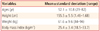
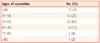
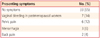
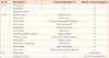
The patient's ages ranged from 29 to 82 years, with a mean of 52.1±10.8 years. The mean tumor size was 5.6 ± 3.5 cm (range, 1-15 cm). Forty six patients (90%) had their tumors in the uterine corpus, and 3 patients (6%) had their tumors in the broad ligament. One in the cervix and one retroperitoneal tumor beneath posterior cul-de-sac were indentified. All patients underwent ultrasound, computed tomography (CT) or magnetic resonance imaging (MRI). Before surgery, 7 patients (14%) were diagnosed with lipoleiomyoma and 8 patients (16%) were diagnosed with ovarian tumors. No tumor was reported to have any cytological atypia, necrosis or other degenerative changes. There were no recurrence or fatalities related to tumor.
Conclusion
Lipoleiomyoma is a rare, benign, uterine neoplasm that requires no treatment when asymptomatic. The fatty nature of the tumor is valuable in preoperative diagnosis with CT and MRI. But the extrauterine lipoleiomyoma including broad ligament can especially resemble ovarian teratoma. Pathological examination of the surgically excised tumor confirmed the diagnosis.
Figures and Tables

References
1. Wang X, Kumar D, Seidman JD. Uterine lipoleiomyomas: a clinicopathologic study of 50 cases. Int J Gynecol Pathol. 2006. 25:239–242.
2. Aung T, Goto M, Nomoto M, Kitajima S, Douchi T, Yoshinaga M, et al. Uterine lipoleiomyoma: a histopathological review of 17 cases. Pathol Int. 2004. 54:751–758.
3. Scurry J, Hack M. Leiomyosarcoma arising in a lipoleiomyoma. Gynecol Oncol. 1990. 39:381–383.
4. Loffroy R, Nezzal N, Mejean N, Sagot P, Krausé D. Lipoleiomyoma of the uterus: imaging features. Gynecol Obstet Invest. 2008. 66:73–75.
5. Ekici E, Vicdan K. Uterine myolipoma: diagnosis by ultrasound. Int J Gynaecol Obstet. 1993. 42:167–171.
6. Prieto A, Crespo C, Pardo A, Docal I, Calzada J, Alonso P. Uterine lipoleiomyomas: US and CT findings. Abdom Imaging. 2000. 25:655–657.
7. Aizenstein R, Wilbur AC, Aizenstein S. CT and MRI of uterine lipoleiomyoma. Gynecol Oncol. 1991. 40:274–276.
8. Sieiński W. Lipomatous neometaplasia of the uterus. Report of 11 cases with discussion of histogenesis and pathogenesis. Int J Gynecol Pathol. 1989. 8:357–363.
9. Tsushima Y, Kita T, Yamamoto K. Uterine lipoleiomyoma: MRI, CT and ultrasonographic findings. Br J Radiol. 1997. 70:1068–1070.
10. Shintaku M. Lipoleiomyomatous tumors of the uterus: a heterogeneous group? Histopathological study of five cases. Pathol Int. 1996. 46:498–502.
11. Oppenheimer DA, Carroll BA, Young SW. Lipoleiomyoma of the uterus. J Comput Assist Tomogr. 1982. 6:640–642.
 XML Download