

 PDF
PDF ePub
ePub Citation
 Print
Print


Abstract
Primary fallopian tube carcinoma is one of the rarest gynecological malignancies, accounting for 0.18% to 1.6% of all malignant neoplasms of the female reproductive tract. Preoperative diagnosis was difficult due to nonspecific symptoms and signs. This case of primary tubal cancer was diagnosed preoperatively on the basis of ultrasonography, computed tomography and magnetic resonance imaging. We have experienced a case of primary fallopian tube carcinoma before operation and so report with brief review of the literature.
Figures and Tables
Fig. 1
Ultrasonographic finding of the primary fallopian tube cancer. It shows a bilocular cystic mass with intramural nodule, measuring 65×36 mm.

Fig. 2
Pelvic computed tomography finding of the primary tube cancer. Bilateral tubular shaped cystic masses with a nodular enhancing solid mass in the cystic mass of the right one (right, measuring 60×43×55 mm; left, 53×29 mm).
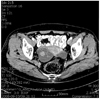
Fig. 3
Pelvic magnetic resonance imaging finding of the primary tubal cancer. Bilateral tubular shaped cystic masses with high S1 on T2W1 with an enhancing solid mural nodule in the Rt. cystic mass, measuring 56×45×45 mm on right and 50×24×50 mm on left.
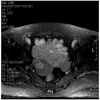
Fig. 4
(A) The right fallopian tube is dilated due to hydrosalpinx the right ovary is grossly unremarkable. (B) Right fallopian tube reveal whitish fiable solid mass (1.8×1.8×1.8 cm) in lumen.

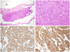
Fig. 5
(A) The lower power view of microscopic finding of solid tumor shows intraluminal growth of fallopian tube with partially infiltrate into the myometrium (H&E, ×40). (B) The high power view of solid tumor reveals no papillary or glandular configuration with prominent nuclear pleomorphism and increased atypical mitoses (H&E, ×200). (C) The immunohistochemical stain of WT-1 reveals strong nuclear expression (×100). (D) The immunohistochemical stain of cancer antigen 125 demonstrates diffusely strong positivity of membranous pattern (×100).
References
1. Rosenblatt KA, Weiss NS, Schwartz SM. Incidence of malignant fallopian tube tumors. Gynecol Oncol. 1989. 35:236–239.
2. Dodson MG, Ford JH Jr, Averette HE. Clinical aspects of fallopian tube carcinoma. Obstet Gynecol. 1970. 36:935–939.
3. Sedlis A. Primary carcinoma of the fallopian tube. Obstet Gynecol Surv. 1961. 16:209–226.
4. Johnston GA Jr. Primary malignancy of the fallopian tube: a clinical review of 13 cases. J Surg Oncol. 1983. 24:304–309.
5. Lootsma-Miklosova E, Aalders JG, Willemse PH, de Bruijn HW. Levels of CA 125 in patients with recurrent carcinoma of the fallopian tube: two case histories. Eur J Obstet Gynecol Reprod Biol. 1987. 24:231–235.
6. Kurjak A, Kupesic S, Ilijas M, Sparac V, Kosuta D. Preoperative diagnosis of primary fallopian tube carcinoma. Gynecol Oncol. 1998. 68:29–34.
7. Kawakami S, Togashi K, Kimura I, Nakano Y, Koshiyama M, Takakura K, et al. Primary malignant tumor of the fallopian tube: appearance at CT and MR imaging. Radiology. 1993. 186:503–508.
8. Kahng YR, Kim JK, Cho KS. Primary malignant tumor of the fallopian tube: CT and MR features. J Korean Radiol Soc. 2001. 45:393–397.
9. Subramanyam BR, Raghavendra BN, Whalen CA, Yee J. Ultrasonic features of fallopian tube carcinoma. J Ultrasound Med. 1984. 3:391–393.
10. Roberts JA, Lifshitz S. Primary adenocarcinoma of the fallopian tube. Gynecol Oncol. 1982. 13:301–308.
11. Hu CY, Taymor ML, Hertig AT. Primary carcinoma of the fallopian tube. Am J Obstet Gynecol. 1950. 59:58–67.
12. Kim TS, Jang HS, Han DG, Kim SL, Choi YC. A case of primary adenosquamous carcinoma of the fallopian tube. Korean J Obstet Gynecol. 1994. 37:1865–1871.
13. Oh YS, Yi SW, Huh CY, Kim SB. Two cases of primary carcinoma of the fallopian tube. Korean J Obstet Gynecol. 1999. 42:1849–1853.
14. Benedet JL, Bender H, Jones H 3rd, Ngan HY, Pecorelli S. FIGO Committee on Gynecologic Oncology. FIGO staging classifications and clinical practice guidelines in the management of gynecologic cancers. Int J Gynaecol Obstet. 2000. 70:209–262.
15. Jung YJ, Chi KS, Kim JS, Kim KW, Kim DG, Yang HS, et al. 3 cases of primary tubal cancer incidentally diagnosed after benign gynecologic operation. Korean J Obstet Gynecol. 2006. 49:1779–1787.
 XML Download