

 PDF
PDF ePub
ePub Citation
 Print
Print


Congenital cataracts account for 30% of congenital eye malformations and have an estimated incidence of 2.3 per 10,000 live births [1]. These cataracts account for 10% of blindness cases in childhood [2]. Congenital cataracts can cause loss of vision unless surgery is performed immediately after birth [3,4]. Therefore, early diagnosis, in particular prenatal diagnosis, is most important, especially because this condition is difficult to diagnose postnatally. Recent advances in obstetric ultrasonography have enabled the diagnosis of fetal eye anomalies such as these. We report a rare Korean case wherein obstetric ultrasonography is used to detect idiopathic congenital cataracts in an otherwise normal fetus.
Case Report
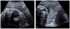
A 28-year-old primigravida who was transferred from a local clinic to our institution underwent routine ultrasonography at 28 weeks and 4 days of gestation. The ultrasonography showed abnormal echogenic spots on both eyes of the fetus (Fig. 1). Both the women and her husband did not have any family history of congenital or metabolic disorders. The patient's obstetric history and medical history was unremarkable, except for a hemorrhoidectomy that was performed 2 years before. Her first-trimester serum screening indicated she was at low risk for aneuploidy. Obstetric ultrasonography performed at 32 weeks of gestation showed highly echogenic areas on both lenses of the fetus. The ultrasonography findings were indicative of congenital cataracts on both eyes. The fetus otherwise seemed normal. The placenta was anterior and not low, and the amniotic fluid volume was normal. Antenatal diagnosis of congenital infections (toxoplasmosis, other infections, rubella, cytomegalovirus [CMV], TORCH; herpes simplex virus) was recommended even though no history of prenatal infection was observed. However, the patient refused diagnostic amniocentesis. Her serum immunoglobin M (IgM) test for rubella yielded negative results, and her serum IgG level for rubella was 6.9 IU/mL at 32 weeks of gestation. Follow-up obstetric ultrasonography performed at 36 weeks and 3 days gestation indicated similar findings, except for the detection of fetal growth restriction. Subsequent obstetric ultrasonography confirmed the presence of hyperechogenic areas on the lenses in both eyes and fetal growth restriction. Although fetal growth restriction was observed, her nonstress test pattern was reactive. At 38 weeks and 3 days of gestation, laboratory test indicated that the patient's serum was non-reactive for venereal disease and negative for antibodies against human immunodeficiency virus.
The patient was hospitalized, and labor was induced at 39 weeks and 1 day of gestation due to fetal growth restriction. On the day of admission, her white blood cell count, hemoglobin level, hematocrit, and platelet count were 7,770 cells/µL, 12.4 g/dL, 36.4%, and 220×103 cells/µL, respectively. The induction of labor progressed poorly, and contraction stress tests revealed fetal distress. Therefore, an emergency Cesarean section was performed, and the infant was delivered at 39 weeks and 2 days of gestation. The female neonate had a birth weight of 2,780 g and a length of 48 cm, and her physical examination did not indicate any abnormalities. An ophthalmologic examination showed normal light reflex of both eyes. Although both corneas were clear, a thick anterior subcapsular opacity with surrounding opacity was observed, indicative of congenital cataracts in both the eyes. Because of the opacity of both lenses, the posterior wall of the eyes could not be visualized; therefore, a B-scan was performed to examine the posterior wall. The ocular ultrasonography B-scan provided non-specific results, except for the cataract finding.
The infant's initial white blood cell count, hemoglobin level, hematocrit, and platelet count were 29,180 cells/µL, 17.1 g/dL, 49.3%, and 393×103 cells/µL, respectively. The infantogram, brain, and kidney scans were all normal. An echocardiogram showed that the diameter of the patent ductus arteriosus was 2 mm. The infant's metabolic disorder test results were all normal, and her TORCH titers were negative, except for the presence of CMV IgG. A maternal immunologic test for CMV showed positive results for CMV IgG suggesting that maternal CMV IgG had been passed to the infant via the placenta. No other abnormalities were detected in the infant during any subsequent examination. The cause of the congenital cataracts remains undetermined. Regardless, lensectomies were performed in both eyes when the infant was 16 days old for improving vision.
Discussion
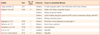
A cataract is any opacity of the lens sufficient to cause visual impairment. The estimated incidence of congenital cataracts is of approximately 2.3 per 10,000 live births [1]. Congenital cataracts have various etiologies, with the most common cause being inheritance as an autosomal dominant trait [5]. Other causes include genetic abnormalities (chromosomal trisomies, chromosomal deletions, and autosomal recessive inheritance), metabolic disorder (diabetes, galactosemia, hypoglycemia, and galactokinase deficiency), a broad spectrum of syndromes (Lowe syndrome, Alport syndrome, Hallermann-Streiff-Francois syndrome, Smith-Lemli-Opitz syndrome, Stickler syndrome, Marinesco-Sjögren syndrome, Zellweger syndrome, myotonic dystrophy, Cockayne syndrome, incontinentia pigmenti, and ichthyosis), TORCH infections, as well as trauma, and radiation exposure [6-8]. However, most congenital cataracts are idiopathic. Based on literature reviews available on PubMed since the year 2000, Table 1 was constructed to list the causes of prenatally diagnosed congenital cataracts and provide basic information about each reported case. We searched the literature in Korea for cases similar to our case. There have been 2 reported cases of idiopathic congenital cataract, including our case. The cause of congenital cataract in our case was also idiopathic.
For better lifelong visual outcome, the timing of surgery in congenital cataract is important. Unilateral cataract surgeries performed within 6 weeks of birth and bilateral cataract surgeries performed within 10 weeks of birth have been reported to result in good outcomes [3,14]. Ocular examination of neonates is difficult, and ocular problems in infants can be easily missed. Consequently, timely diagnosis and corrective surgery may be difficult in infants. Prenatal diagnosis of congenital cataract can resolve this problem. Advances in obstetric ultrasonography have enabled prenatal diagnosis of ocular anomalies. Transvaginal ultrasonography also enables the detection of fetal cataracts in early pregnancy. Normal obstetric ultrasonographic findings for the fetal lens include a smooth echogenic ring with a diameter of approximately 4 mm at 18 weeks of gestation and of approximately 7 mm at term with hypoechogenic content [4]. Obstetric ultrasonography findings of congenital cataracts include thick, irregular, or crenated hyperechogenic borders; a cluster of hyperechogenic material; or homogeneous opacity [15]. Obstetricians should screen for these abnormalities and carefully examine ocular anomalies for timely treatment of congenital cataracts. Thorough ultrasonographic examinations for prenatal cataracts are indicated for patients who have a family history of this condition, for those who are suspected of having a TORCH infection or fetal anomaly, or for those who are exposed to radiation. Early diagnosis of congenital cataracts in a fetus with severe congenital malformations allows the parents to be better prepared for the disease. Additionally, with early diagnosis, the physician may easily counsel the mother.
 XML Download