

 PDF
PDF ePub
ePub Citation
 Print
Print


The mucoepidermoid carcinoma (MEC) is an epithelial malignant tumor that Stewart et al. [1] first described as a salivary gland tumor. MEC is very common in salivary gland, but is rare in the cervix. It composes 30% of malignant tumors rising in major or minor salivary glands [2]. And other organs such as lung [3] and esophagus are also known as primary tumor site. We experienced a case of primary MEC in the cervix, and reported the case in this review as the first report in Korea.
Case Report
A 44-year-old female visited to Samsung Medical Center due to abnormal finding in cervical biopsy on routine screening. The pathologic finding of punch biopsy was MEC. She had no specific symptom. Under gynecologic examination, about 2 cm-sized cancerous mass was noted in anterior lip of cervix. The mass was located only in cervix not extended to vagina or parametrium. Abdomen-pelvis magnetic resonance imaging (MRI) showed 2.6×1.8×0.9 cm-sized cancerous mass arising from anterior lip of uterine cervix with possible invasion to upper vagina (Fig. 1). The radical hysterectomy, right salpingoophorectomy and pelvic lymphadenectomy by laparoscopic method were performed.
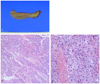
Grossly, there was a relatively well demarcated mass, measuring 2 ×1.7 ×1.5 cm. It was confined within the cervix (Fig. 2). Histopathological examination showed that tumor was composed of squamous, intermediate and mucin secreting cells that together formed a variety of patterns with necrotic background. Mucin secreting cells showed clear vacuolated cytoplasm forming solid sheet pattern. The malignant cells had prominent nucleoli and pleomorphic nuclei suggestive of high grade tumor. There was no metastasis to regional lymph node. Both resection margins were tumor free. Therefore, the patient was diagnosed as cervical MEC, International Federation of Gynecology and Obstetrics (FIGO) stage IB1 without any risk factors. We had decided to closely observe without adjuvant therapy. She complained left flank pain and bowel habit change 4 months after the primary surgical treatment. After evaluation, about 8.5 cm-sized heterogeneous mass were found in left side of vaginl stump and, near to this cancerous mass, 2.3 cm-sized lobulated mass were also noted. Those masses had invaded into left distal ureter causing hydronephroureterosis. Multiple metastatic lymph nodes were noted in para-aortic area, pelvic cavity and mesocolon. We planned to start concurrent chemoradiation (CCRT) with paclitaxel and carboplatin. During CCRT, recurrent vaginal mass showed interval improvement with decreasing tumor size. One month after CCRT, the size of previous mentioned cancerous mass in vaginal stump increased and the mass invaded into the left wall of pelvis and posterior wall of urinary bladder. Radiotherapy was not indicated for treating recurred masses. We started the palliative chemotherapy with irinotecan and cisplatin. Despite the effort of our management, recurred mass in vaginal stump was aggravated and new masses in the right pelvic cavity, left common iliac lymph node and right perirectal lymph node were noted. The multiple metastatic tumors were very aggressive and had resistance in chemotherapy. After all, she expired after 19 months of primary surgical treatment.
Discussion
MEC in the cervix is included in invasive carcinoma, which is similar to its salivary counterpart. There are several previous reports about cervical MEC [4,5]. The morphology is similar between cervical and salivary gland MEC. MEC of cervix is a controversial entity. MEC is a kind of adenosquamous carcinoma and its frequency in cervical cancer is not documented [6]. But recent reports demonstrate that the tumor diagnosed as MEC according to morphologic criteria have genetic mutations involving the genes characteristically rearranged in the salivary glands. Those reports suggested that the cervical MEC is unique entity distinct from cervical adenosquamous carcinoma [7]. Thelmo et al. [5] mentioned that mucoepidermoid carcinoma was more aggressive cancer. Twelve women were followed for 2 to 15 years. Three women died within one year. All patients had lymph node and vascular metastases. Several other papers have reported that MEC of cervix also has different clinical manifestation [5], including frequent metastasis and sensitivity to radiotherapy compared with adenosquamous carcinoma of cervix [8]. But, MEC of cervix is not divided from cervical adenosquamous carcinoma. Since the diagnosis of cervical MEC bring therapeutic significance, the morphologic criteria to distinguish cervical MEC from adenosquamous carcinoma is important.
There is no standard treatment in cervical MEC because of a controversial entity and limited data. In general, cervical cancer treatment is different in each stage. In earlier stage, surgical resection is a choice. According to histopathologic risk factors, adjuvant radiotherapy or chemotherapy is needed.
In conclusion, we report a very rare case of MEC in the cervix and this is the first case reported in Korea. There is few report of this entity in cervix and treatment strategy. This case was aggressive in biological behaviors. MEC can present widely diverse behavior based on variety histological characteristics.

 XML Download