

 PDF
PDF ePub
ePub Citation
 Print
Print


Abstract
Objective
Papillary tumor is known to be evaluated the histology through complete resection because of difficulty to distinguish between benign tumor and malignant tumor through core needle biopsy. We introduced clinical experience for better diagnosis of papillary tumor through ultrasound-guided vacuum-assisted resection (Mammotome).
Methods
We carried out core needle biopsy and Mammotome for 25 patients diagnosed to papillary tumor and found out the pathologic results.
Figures and Tables
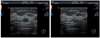 | Fig. 1Core needle biopy procedure, this sonogram of breast shows the core needle penestrating the central portion of the breast mass avoiding vessels via color Dppler.
|
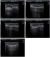 | Fig. 2Mammotome breast mass resection procedure, this each sonogram of mammotome procedure show each step sequentially.
|
| Fig. 3Sonographic features of papillary tumor of breast. (A) Intraductal mass. (B) Intracystic mass. (C) Solid mass with peripheral anechoic rim. (D) Extraductal mass adjacent to the dilated duct. (E) Mixed type (intraductdal+extraductal).
|
References
1. Korea Central Cancer Registry. Annual report of cancer statistics in Korea 2008 [Internet]. c2011. cited 2011 Mar 2. Goyang: National Cancer Center;Available from: http://www.cancer.go.kr/ncic/cics_f/01/011/index.html.
2. American College of Radiology. Breast imaging reporting and data system: breast imaging atlas: mammography, breast ultrasound, breast MR imaging. 2003. 4th ed. Reston (VA): American College of Radiology.
3. Page DL, Anderson TJ. Diagnostic histopathology of the breast. 1987. London: Churchill Livingstone.
4. Raju UB, Lee MW, Zarbo RJ, Crissman JD. Papillary neoplasia of the breast: immunohistochemically defined myoepithelial cells in the diagnosis of benign and malignant papillary breast neoplasms. Mod Pathol. 1989. 2:569–576.
5. Han BK, Choe YH, Ko YH, Yang JH, Nam SJ. Benign papillary lesions of the breast: sonographic-pathologic correlation. J Ultrasound Med. 1999. 18:217–223.
6. Park JH, Bae JS, Suh YJ, Park WC, Song BJ, Kim JS, et al. Clinicopathologic features of the papillary breast lesions diagnosed on ultrasonography-guided core needle biopsy. J Breast Cancer. 2007. 10:269–272.
7. Ra YM, Sohn JS, Kim KW, Lee JU, Park HD, Choi IS, et al. Clinicopathologic review of the intraductal papilloma of breast. J Breast Cancer. 2010. 13:31–36.
8. Ribeiro-Silva A, Zambelli Ramalho LN, Britto Garcia S, Zucoloto S. The relationship between p63 and p53 expression in normal and neoplastic breast tissue. Arch Pathol Lab Med. 2003. 127:336–340.
9. Werling RW, Hwang H, Yaziji H, Gown AM. Immunohistochemical distinction of invasive from noninvasive breast lesions: a comparative study of p63 versus calponin and smooth muscle myosin heavy chain. Am J Surg Pathol. 2003. 27:82–90.
10. Kesse-Adu R, Shousha S. Myoepithelial markers are expressed in at least 29% of oestrogen receptor negative invasive breast carcinoma. Mod Pathol. 2004. 17:646–652.
11. Moriya T, Kasajima A, Ishida K, Kariya Y, Akahira J, Endoh M, et al. New trends of immunohistochemistry for making differential diagnosis of breast lesions. Med Mol Morphol. 2006. 39:8–13.
12. Liberman L, Tornos C, Huzjan R, Bartella L, Morris EA, Dershaw DD. Is surgical excision warranted after benign, concordant diagnosis of papilloma at percutaneous breast biopsy? AJR Am J Roentgenol. 2006. 186:1328–1334.
13. Lam WW, Chu WC, Tang AP, Tse G, Ma TK. Role of radiologic features in the management of papillary lesions of the breast. AJR Am J Roentgenol. 2006. 186:1322–1327.
14. Sohn V, Keylock J, Arthurs Z, Wilson A, Herbert G, Perry J, et al. Breast papillomas in the era of percutaneous needle biopsy. Ann Surg Oncol. 2007. 14:2979–2984.
15. Jung SY, Kang HS, Kwon Y, Min SY, Kim EA, Ko KL, et al. Risk factors for malignancy in benign papillomas of the breast on core needle biopsy. World J Surg. 2010. 34:261–265.
16. Lewis JT, Hartmann LC, Vierkant RA, Maloney SD, Shane Pankratz V, Allers TM, et al. An analysis of breast cancer risk in women with single, multiple, and atypical papilloma. Am J Surg Pathol. 2006. 30:665–672.
17. Carder PJ, Garvican J, Haigh I, Liston JC. Needle core biopsy can reliably distinguish between benign and malignant papillary lesions of the breast. Histopathology. 2005. 46:320–327.
18. Hoda SA, Rosen PP. Observations on the pathologic diagnosis of selected unusual lesions in needle core biopsies of breast. Breast J. 2004. 10:522–527.
19. Valdes EK, Tartter PI, Genelus-Dominique E, Guilbaud DA, Rosenbaum-Smith S, Estabrook A. Significance of papillary lesions at percutaneous breast biopsy. Ann Surg Oncol. 2006. 13:480–482.
20. Ko ES, Cho N, Yang SK, Kim DY, Moon WK. Papillary lesions of the breast: comparison of the US-guided 14-gauge automated gun method and the 11-gauge directional vacuum-assisted biopsy method. J Korean Radiol Soc. 2006. 54:537–541.
21. Mercado CL, Hamele-Bena D, Singer C, Koenigsberg T, Pile-Spellman E, Higgins H, et al. Papillary lesions of the breast: evaluation with stereotactic directional vacuum-assisted biopsy. Radiology. 2001. 221:650–655.
22. Rosen EL, Bentley RC, Baker JA, Soo MS. Imaging-guided core needle biopsy of papillary lesions of the breast. AJR Am J Roentgenol. 2002. 179:1185–1192.
23. Mercado CL, Hamele-Bena D, Oken SM, Singer CI, Cangiarella J. Papillary lesions of the breast at percutaneous core-needle biopsy. Radiology. 2006. 238:801–808.
24. Wei H, Jiayi F, Qinping Z, Junyi S, Yuan S, Li L, et al. Ultrasound-guided vacuum-assisted breast biopsy system for diagnosis and minimally invasive excision of intraductal papilloma without nipple discharge. World J Surg. 2009. 33:2579–2581.
25. Norton LW, Pearlman NW. Needle localization breast biopsy: accuracy versus cost. Am J Surg. 1988. 156:13B–15B.
26. Fine RE, Whitworth PW, Kim JA, Harness JK, Boyd BA, Burak WE Jr. Low-risk palpable breast masses removed using a vacuum-assisted hand-held device. Am J Surg. 2003. 186:362–367.
27. Kim MJ, Kim EK, Lee JY, Youk JH, Park BW, Kim SI, et al. Breast lesions with imaging-histologic discordance during US-guided 14G automated core biopsy: can the directional vacuum-assisted removal replace the surgical excision? Initial findings. Eur Radiol. 2007. 17:2376–2383.
28. Parker SH, Klaus AJ, McWey PJ, Schilling KJ, Cupples TE, Duchesne N, et al. Sonographically guided directional vacuum-assisted breast biopsy using a handheld device. AJR Am J Roentgenol. 2001. 177:405–408.
29. Perez-Fuentes JA, Longobardi IR, Acosta VF, Marin CE, Liberman L. Sonographically guided directional vacuum-assisted breast biopsy: preliminary experience in Venezuela. AJR Am J Roentgenol. 2001. 177:1459–1463.
30. March DE, Coughlin BF, Barham RB, Goulart RA, Klein SV, Bur ME, et al. Breast masses: removal of all US evidence during biopsy by using a handheld vacuum-assisted device: initial experience. Radiology. 2003. 227:549–555.
 XML Download