

 PDF
PDF ePub
ePub Citation
 Print
Print


Two placentas are rare in pregnancies, including succenturiate placental [1]. Two placentas with fused umbilical cord forming 3 vessels cord at the fetal end which has its own insertion site to each placental disc is an extremely rare case in a singleton pregnancy. In this case, we observed two placentas with fused umbilical cord with an episode of vanishing twin syndrome and there seems to be a strong relationship between these two events. Here, we report two placents in sington pregnancy with fused umbilical cord in the pregnant woman of vanishing twin.
Case Report
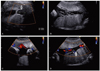
A 37-year-old woman, gravid 0, para 0, visited emergency room with an episode of vaginal bleeding, diagnosed as threatened abortion with 1.93×1.71 cm sized subchorionic hematoma. The fetus was 8 weeks and 5 days sized which was resulted from in vitro fertilization-embryo transfer (IVF-ET) and there was 2.23×1.62 cm (about 6 weeks size) gestational sac like lesion at uterine fundus (Fig. 1A), Follow-up ultrasonic evaluation revealed previously found gestational sac like lesion which contained no embryo with subchorionic hematoma and this appeared to be vanishing twin syndrome. No additional subchorionic hematoma and empty gestational sac were found at 21 gestational week, however, the placenta appeared equally divided into anterior and posterior lobes with umbilical cord containing 3 vessels (Fig. 1B). Closer inspection with follow-up ultrasonic exam confirmed two placentas-anterior and posterior, respectively-and the cord Doppler systolic/diastolic (S/D) ratio remained in the normal range 1.9 and 2.24, respectively. Among the two placentas, it was difficult to differentiate main placenta which supply fetus because of the individual umbilical cord of each placenta and the complexity of two umbilical cords in amniotic cavity (Fig. 1C, 1D).
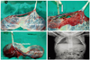
At 40 weeks and 4 days, the patient delivered a viable female infant weighing 3,900 g via Cesarean section with diagnosis of suspicious cephalopelvic disproportion and an Apgar score was 9/10 at 1 and 5 minutes. Inspection of the postpartum placentas, membranes, and cord confirmed ultrasound findings. The placenta consists of two placental discs-13×16×3 cm sized main disc and 13×12×2.5 cm sized side disc, respectively-and there were 2 cord insertions, 1 into each placenta (Fig. 2A). The cord at each of the placental disc had marginal insertion site and main placental disc cord had 2 arteries with one vein (3 vessel cord) whereas side placental disc cord had one artery with one vein (2 vessel cord) (Fig. 2B, 2C). The umbilical cord had total length of 24.5 cm and the umbilical cord of the side placental disc was fused at the insertion site of the main umbilical cord forming normal 3 vessel cord at the fetal end. The fused umbilical cord measured 11 cm in length and 0.8 cm in diameter.
After delivery, the baby was taken whole body X-ray and abdominal ultrasound to exclude fetus in fetus and 2-D echocardiography was also taken to exclude cardiac input overloads resulted from two placentas. No abnormalities were found in the evaluation of the infant (Fig. 2D).
Discussion
Two placentas are rare in pregnancies, including succenturiate placental [1]. Two placentas with fused umbilical cord forming 3 vessels cord at the fetal end which has its own insertion site to each placental disc is an extremely rare case in a singleton pregnancy. Moreover, this present case describes two placentas with fused umbilical cord with an episode of vanishing twin syndrome and there seems to be a strong relationship between these two events. There was only one case of a duplicated placenta and bifurcated umbilical cord in a singleton pregnancy, but this case was not related with vanishing twin [2]. Therefore, this present case may be the first case which describes two placentas with fused umbilical cord that is related with vanishing twin.
Vanishing twin syndrome is the identification of a multiple gestation with subsequent disappearance of one or more fetuses. The rate of multiple gestations at conception is higher than the incidence noted at birth [3]. The frequency of multiple gestations is 3.3% to 5.4% at 8 weeks' gestation [2]. Vanishing twin syndrome occurs in 21% to 30% of multiple gestation [4]. This vanishing twin case was resulted from IVF-ET with one normal pregnancy and one gestational sac containing no embryo. A relationship between placenta morphologic features and the superficial implantation and/or inadequate orientation of the blastocyst after IVF has been proposed [5]. Considering the fact that this present case was resulted from IVF-ET, these well-known relationships between placenta morphology and IVF-ET could account for the two placentas in this case.
According to classification of placental morphology, two separate placentas of this case could be considered as succenturiate placenta [6] which provide another possible hypothesis for this case. Presenting on ultrasound as a small section of placental tissue distant to the main placental body, they are thought to represent a form of trophotropism of the placenta. It is believed that the placenta recedes from areas of inadequate blood supply such as fibroids and may experience some proliferation of villi on the other placental margins. Thus, trophotropism may result in a separated, or succenturiate, section of the placenta [2]. Suzuki et al. [7] reported that the incidence of succenturiate lobes of placenta in twin pregnancies was significantly higher than that in singleton pregnancies. Furthermore, in their earlier study with singleton pregnancies, Suzuki and Igarashi [8] also reported the frequency of maternal age >35 years and history of infertility using IVF in patients complicated by succenturiate lobes of placenta were significantly higher than those without succenturiate lobes of placenta. In this present case, vanishing twin syndrome may attribute to this two placentas. However, the diagnosis of succenturiate lobes of placenta requires the additional placental lobe that is much smaller than the largest lobe of placenta macroscopically and the presence of subchorionic vessels between the main placental disk and the accessory lobe confirmed by placental pathologist [2,7]. In this case, the placentas were almost equal in size and placed on both anterior and posterior portion of the uterus. The only connection between the two placental discs was the free-floating, fused umbilical cord without connection of subchorionic vessels. Thus, in this case, a succenturiate lobe could be considered according to the placenta classification, but this special morphology is not match with succenturate lobes of placenta that could be explained true duplicated placentas.
In vanishing twin syndrome, there may be complete reabsorption of a fetus, formation of a fetus papyraceus, or development of a subtle abnormality on the placenta such as a cyst, subchorionic fibrin, or amorphous material [9]. In this case, repeated antenatal ultrasonic exam revealed complete reabsorption of the gestational sac which contained no embryo. During the process of reabsorption, fetus in fetus, which was first described by Meckel, was thought to be ruled out for continuously developing side placental disc and fused umbilical cord. Fetus in fetus is a rare condition in which a malformed parasitic twin was found inside the body of its partner, usually in the abdominal cavity. It represents an aberration of monozygotic diamniotic twinning [10]. We considered the possibility of fetus in fetus with two placentas. However, in this case, no abnormalities were found during the repeated antenatal ultrasound exams and the findings of whole body X-ray and abdominal ultrasound for the neonate were normal. Furthermore, neonatal 2-D Echocardiography of the infant was also normal.
There are several reports which describes bifurcated umbilical cords in monoamniotic twin gestations [11]. A bifurcated umbilical cord in a monoamniotic twin gestation is an event that takes place when separation of the 2 embryos is delayed until just hours before they would have become conjoined on day 13 postfertilization [1,11]. However, to our knowledge, this present case is the first case of two placentas with fused umbilical cord which originated from vanishing twin syndrome in singleton pregnancy and further evaluation of etiology and morphologic features is needed.
 XML Download