

 PDF
PDF ePub
ePub Citation
 Print
Print


Preterm labor is defined as the presence of regular uterine contractions with progressive cervical changes before 37 completed weeks of pregnancy. Spontaneous preterm labor accounts for 40% to 50% of all preterm births [1]. An estimated 13 million babies (-10% of all births) are born preterm each year worldwide, and approximately 1 million of all newborn deaths are due to preterm birth [2].
At present, typical uterine contraction inhibitors (e.g., betamimetics, magnesium sulfate, calcium channel blockers, oxytocin receptor antagonists, and prostaglandin inhibitors) have achieved limited success for short-term management of preterm labor. Although tocolytic agents were superior to placebo at delaying delivery for at least 7 days in a meta-analytic study, none was statistically more effective than placebo at delaying delivery to 37 weeks gestation or at reducing neonatal mortality and morbidity [3]. Women receiving tocolytics for preterm labor require monitoring and intensive support for serious side effects that may affect both the mother and fetus, including chest pain, dyspnea, palpitations, tremors, headaches, hyperglycemia, hypokalemia, and fetal tachycardia [4]. Despite the development of modern neonatal care, infants born prior to 28 weeks of gestation, in whom fetal lung development is premature, exhibit high mortality and morbidity rates [5]. Extending pregnancy by a few more weeks can make a huge difference in neonatal outcomes. Therefore, a new treatment approach that results in long-term prevention of premature uterine contraction is required to lower neonate mortality.
Clostridium botulinum toxin A (BoNT/A) blocks the release of the neurotransmitter acetylcholine (ACh) from presynaptic nerve endings [6]. While it has been primarily shown to affect skeletal muscles, there is much evidence showing that BoNT/A also affects smooth muscles, including the gastrointestinal [7] and detrusor muscle [8]. Recently, two reports supported its effects on the myometrium. In 2003, Garza et al. [9] demonstrated that BoNT/A inhibits the spontaneous uterine activity of pregnant rat myometrium in vitro. In 2009, Burd et al. [10] reported that BoNT/A suppresses oxytocin-induced contractions of pregnant human myometrium in vitro. Currently, BoNT/A is approved by the Food and Drug Administration (FDA) for several clinical indications, including strabismus, blepharospasm, facial nerve VII disorders, cervical dystonia, primary axillary hyperhidrosis, and chronic migraine headaches, and as a cosmetic treatment for glabellar lines. Other uses of BoNT/A that are successful treatments not yet approved by FDA include treatment of achalasia, lower urinary tract dysfunction, chronic anal fissures, various spastic disorders, neuropathies, wound healing, and various cosmetic indications.
The purpose of this study was to evaluate the tocolytic effect of BoNT/A on mifepristone-induced preterm labor in pregnant rats by determining changes in electromyography (EMG) activity of uterine contractility in vivo.
Materials and Methods
1. Animals
Fourteen primiparous Sprague-Dawley rats of approximately 10 weeks of age were supplied by Nara Biotech (Seoul, Korea). The average body weight of the rats was 309 g (range, 245-354 g). Determination of pregnancy was made by the observation of a vaginal plug. Plug observation date was considered to be day 1 of gestation. The pregnant rats were caged singly until the animals arrived in our laboratory on day 16 of gestation. In our laboratory, two rats were housed together in a standard animal cage (420 × 270 ×180 mm). Rats had free access to water and pellets, were housed in a light-controlled room with 13 hours of light (7 AM-8 PM) and 11 hours of darkness, and were maintained at a temperature of 22℃ to 24℃. The animal experimentation described in this study was approved by the CHA University School of Medicine Institutional Animal Care and Use Committee.
2. Anesthesia
The animals were anesthetized for both surgical procedures and EMG recordings. General anesthesia was induced by intramuscular injection of a mixture of 0.06 mL tiletamine plus zolazepam (Zoletil 50, Virbac, Carros, France) and 0.06 mL xylazine (Rompun, Bayer Korea, Seoul, Korea). If a surgical procedure was prolonged or induction of anesthesia was insufficient, a subsequent intramuscular injection was given at one-half the original dose.
3. Drugs and treatments
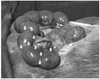
A total of fourteen rats were randomly assigned into two groups. Seven rats were assigned as a sham-operated group (group A), which were treated with normal saline. Seven rats were assigned as an experimental group (group B), which were administered BoNT/A at dose of 20 U. On day 17 of gestation, both uterine horns were exposed through a midline abdominal incision under sterile conditions in all animals. Each rat in the sham-operated group was administered a total volume of 1.2 mL of normal saline on both uterine horns (0.6 mL of normal saline on each uterine horn), regardless of differences in uterine horn size. Experimental group was administered a total volume of 1.2 mL of 20 U BoNT/A (Neuronox, Medy-Tox, Seoul, Korea). Injections were made on the lower portion of each uterine horn, in an area where a large amount of the uterine myometrium is located, and the mesometrium is attached (Fig. 1). Injections were also made in an area between the bulging region of the gestational lumen of both sides and the upper middle portion of the uterine horn. Direct injections into the gestational sac were avoided. Special needles (Johns screw needles, Woorhi Medical, Seoul, Korea), which offer control of needle length from 0 to 7 mm, were used at a needle length of 1 mm. After injections on the uterus, the abdominal wall and skin were closed with continuous running sutures of #3-0 nylon. Preterm labor was induced with mifepristone (RU-486, Ascent Scientific, Cambridge, UK) in all animals [11]. On day 18 of gestation, a single subcutaneous injection of mifepristone (3 mg suspended in 0.2 mL olive oil) was made in the rat buttocks, which resulted in preterm labor within 24 hours.
4. EMG recordings
A Medelec Synergy N2 machine (Oxford Instruments, Oxfordshire, UK) was used to record the EMG activity of uterine contractility. EMG activity was recorded on day 19 (approximately 23 to 24 hours after mifepristone injection). Uterine EMG activity was recorded from the abdominal surface. A number of animal and human studies have shown transabdominal surface measurements of uterine EMG activity to correlate well with intrauterine pressure [12]. All animals were anesthetized for EMG recordings. Abdominal hair was removed with 80% thioglycolic acid cream. A pair of disposable surface pickup electrodes was placed approximately 10 to 15 mm apart on the abdominal wall, overlaying the uterine horn. A reference electrode was placed near the chest area to reduce artifacts caused by movements of the chest wall. The band-pass filter was set from 0.5 to 50 Hz. At the end of the EMG recordings (24 hours after mifepristone injection), the number of premature deliveries and pups delivered by each rat were counted, and all rats were euthanized by cervical dislocation under anesthesia.
5. Quantification of data and statistical analyses
All EMG data were recorded and visually analyzed using Medelec Synergy software (ver. 11.1, Oxford Instruments). The duration and latency time (in milliseconds) were analyzed using a vertical cursor. The peak-to-peak amplitude (µV) of each burst was evaluated using a horizontal cursor. The onset of a burst was defined by the amplitude progressively rising to at least twice the baseline electrical activity for a period of at least 5 seconds. All the resultant data is expressed as the mean ± standard error of the mean (SEM). Mean percent change from baseline is expressed as the mean percent change ± SEM% (SEM/mean×100). Statistical comparisons of groups are based on the Wilcoxon rank sum test performed using SAS Enterprise Guide software (ver. 4.1, SAS Institute, Cary, NC, USA). A P-value less than 0.05 indicates a statistically significant difference.
Results
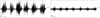
Five of seven rats (71.4%) in the sham-operated group delivered seven pups within 24 hours after treatment with 3 mg mifepristone, whereas two of seven (28.5%) rats in the experimental group delivered two pups prematurely. Uterine EMG activity was characterized by regular, higher amplitude, and longer duration bursts of action potentials in the sham-operated group compared to animals treated with BoNT/A (Fig. 2A). The animals in groups B showed notably reduced uterine electromyographic activity compared to the sham-operated group in terms of decreased amplitude and duration of uterine contractions (Fig. 2B). Increasing the concentration to 20 U of BoNT/A significantly decreased the amplitude of bursts by 45.2% ± 18.4% (P<0.05) compared to that of the sham-operated group (Fig. 3A). The total duration of uterine contractions for these groups was significantly decreased by 51.7% ± 7.9% (P<0.01), respectively (Fig. 3B). The frequency of uterine contractions for animals in group B was decreased by 5.6% ± 16.3% (P=0.4) compared to the sham-operated group (Fig. 3C).
Discussion
To our knowledge, this is the first in vivo study showing that BoNT/A not only inhibits uterine contractions, but also delays rat delivery of pups in mifepristone-induced preterm labor. The decrease in uterine activity was mainly caused by a decline in the duration and amplitude, rather than a decline in the frequency, of uterine contractions. At 20 U of BoNT/A, the decrease in uterine activity was prominent in the electromyographic results. Although the frequency of uterine contractions was not statistically significant (P=0.399) in this experiment. A significant decrease in the frequency of uterine contractions may be expected at higher doses of BoNT/A beyond 20 units, as shown by Garza et al. [9], but this would eventually reach the lethal/toxic threshold of BoNT/A (IM LD50, 114.1±11.5 U/kg; unpublished raw data, Medy-Tox, Seoul, Korea) in vivo. Based on visual observations during EMG recording, the experimental animals treated with BoNT/A showed neither changes in movement or behavior nor excessive vaginal bleeding, except for a small amount of brownish discharge or blood at the lips of the vulva, compared to the sham-operated animals during labor.
BoNT/A is labeled as a pregnancy risk category C drug by the FDA [6], which means that well-controlled human studies are lacking and safety of this drug for use during pregnancy has not been established. Therefore, we do not know whether its use constitutes a chance of fetal harm. In one report in which 16 pregnant women were treated with BoNT/A, no adverse effects on the fetus or mother were observed, except one mother, with a prior history of spontaneous abortions suffered a miscarriage [13]. Uncomplicated full-term pregnancies of four women, who received regular Botox treatments for cervical dystonia, have also been reported [14]. Furthermore, to date, no adverse effects of BoNT/A to either the mother or fetus during pregnancy have been reported. The molecular weight of BoNT is 150 kDa; therefore, it is unlikely that BoNT passes through the placental membrane [13]. However, it is unknown whether an active transport mechanism exists, so potential effects on the fetus should not be ignored.
No clinical trials for Clostridium botulinum toxin type A for inhibition of uterine contractions during preterm labor have been conducted to date. Therefore, we do not know the optimal dose of BoNT/A on the human uterus for prevention of preterm labor. In a meta-analysis of patients with overactive bladder syndrome treated with BoNT/A, lower doses of botulinum toxin (100 U) appeared to elicit beneficial effects; however, larger doses (300 U) were found to be more effective and longer lasting but had more side effects, such as urinary retention and urinary tract infection [15]. If BoNT/A were to be used during pregnancy in humans, lower doses (<100 U) would be recommended, because higher doses may cause unwanted side effects, such as uterine atony and unnecessarily persistent inhibition of uterine contractions. Administration of lower doses of botulinum toxin would provide ongoing suppression of uterine contractions to maintain pregnancy with potentially fewer side effects. In addition, if stronger suppression of uterine contractions is necessary to prevent preterm labor, concomitant administration of a current tocolytic agent, such as betamimetics, and BoNT/A may yield a stronger synergistic inhibition of uterine contractions. In regard to the method of administration, the use of ultrasonography with color-flow Doppler as a guide allows the entrance of a needle (18 gauge spinal needle) and delivery of BoNT/A into the muscular layers of the fundus, avoiding the vascular structures of the uterus, as the mechanical activity of the uterus is initiated in the fundus during normal labor [16].
Clostridium botulinum neurotoxin consists of a zinc-dependent protease light chain (50 kDa) and a membrane-binding heavy chain (100 kDa), which are joined by a disulfide bond [6]. There are seven BoNT serotypes (A, B, C1, D, E, F, and G), defined by differences in the light chains and result in different muscle-affecting properties. BoNT/A and BoNT/E cleave a synaptosome-associated protein of 25 kDa (SNAP-25), while BoNT/B, /D, /F, and /G cleave vesicle-associated membrane protein or synaptobrevin [6]. BoNT/C1 is reported to cleave both syntaxin and SNAP-25, which are essential for vesicle fusion at presynaptic terminals, due to their ability to form a SNARE complex; their cleavage blocks membrane fusion of presynaptic vesicles containing ACh [6,17]. Consequently, inhibition of such neurotransmitter release from presynaptic neurons results in muscle paralysis. Until now, the mechanisms explaining BoNT/A inhibition of uterine myometrial contractions have been unknown. Unlike skeletal muscles, uterine smooth muscles are affected by α- and β-adrenergic receptors [18]. There is also evidence showing that uterine smooth muscle is affected by cholinergic nerves that play important roles in stimulating contraction of myometrium and vasculature [19,20]. Since botulinum toxin blocks cholinergic transmission by inhibiting ACh release, we may postulate that the effect of BoNT/A on cholinergic neurons innervating the uterus is likely the cause of reduced uterine contractions in pregnant rats. Such a view accords well with indirect studies of smooth muscles such as the sphincter of Oddi [21] and the pyloric sphincter [22]. In regards to the pyloric sphincter study, it has been suggested that higher concentrations of botulinum toxin may have a direct inhibitory effect on smooth muscle contractility by acting on smooth muscle muscarinic receptors [22]. Such experimental observations were further supported by a study of the immunohistochemical expression of muscarinic receptors in patients of detrusor overactivity treated with BoNT/A compared with controls. Specifically, it was shown that these patients not only had an improvement in urgency after treatment, but also a significant decline in M1 and M3 receptor immunoreactivity [23]. Interestingly, studies of the pharmacological characteristics of muscarinic receptors in rat and mouse isolated myometrium using selective muscarinic antagonists indicated that the muscarinic M3 receptors are a major mediator of uterine contraction [24,25]. Further exploration is needed to determine the correlation of expression levels of these receptors with BoNT/A in the uterus during pregnancy. These efforts will guide us to gain a better understanding and preventing of preterm labor.
 XML Download