

 PDF
PDF ePub
ePub Citation
 Print
Print


Heterotopic pregnancy (HP) is a rare complication of pregnancy in which intra-uterine and extra-uterine gestation occur simultaneously. The prevalence rate of HP is as rare as 1 in 30,000 natural gestations but increases to 1 in 100 pregnancies in women who have undergone an assisted reproductive technology (ART) procedure [1]. The incidence of cornual HP is estimated to be as high as 1 in 3,600 in vitro fertilization (IVF) pregnancies [2]. Cornual HP has a high risk of uterine rupture and is difficult to diagnose early. The main issue in the treatment of a cornual HP is to be as minimally invasive as possible to preserve the developing intrauterine pregnancy.
We report the case of a cornual heterotopic pregnant woman who delivered a healthy baby after a successful laparoscopic cornual resection for a cornual HP that developed after the women underwent IVF-embryo transfer (ET).
Case Report
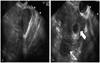
A 28-year-old Korean woman, gravida 2, para 0, underwent IVF-ET because she had undergone two prior salpingectomies for tubal pregnancies at other centers. She was referred to the emergency department because she had experienced an abrupt onset of lower abdominal pain at the 7th gestational week. At initial presentation, her vital signs were stable and physical examination of the abdomen presented lower abdominal tenderness and rebound tenderness. A transvaginal ultrasonogram showed free fluid in the cul-de-sac and two gestational sacs (Fig. 1). One gestational sac was present in the right cornual area and the other was in the uterine cavity. Two separate yolk sacs and fetal parts were noted in each gestational sacs, but there were no fetal heart tones. We performed emergent laparoscopic surgery including right cornual resection.
To minimize exposure of the fetus to anesthetic agents, general anesthesia was started after completion of the preparations of all laparoscopic surgical instruments and operation-related procedures. After the trocar was inserted, carbon dioxide pressure was maintained at 10 to 12 mm Hg or less.
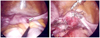
The operative findings were about 500 mL of free blood in the culde-sac, a partially ruptured right cornual mass, and some pelvic adhesions (Fig. 2A).
For cornual wedge resection, a circumferential incision was made 1 to 2 cm above the base of the cornual pregnancy using harmonic shears (Ultracision Harmonic Scalpel, Ethicon Endo-Surgery, Cincinnati, OH, USA). We sutured using extracorporeal, interrupted, sutures of one or two layers using 1-0 and 2-0 polyglactin 910 sutures (Vicryl, Ethicon Inc., Somerville, NJ, USA) (Fig. 2B).
The operating time was 40 minutes, which included the time from skin incision to the time of closure of all port sites. She was transfused with three pints of packed red blood cells during surgery. Antibiotics and analgesic agents were not used before or after the operation. The final histopathological diagnosis was compatible with cornual pregnancy. There were no intra-operative or postoperative complications. On postoperative day 4, a normal fetal heart tone in the endometrial gestational sac was noted and the patient was discharged without any obstetric or surgery-related complications.
The woman delivered a healthy baby of 3,340 g via Caesarean section due to failure to progress at term.
Discussion
The risk factors for HP are endometriosis, periadnexal adhesion, tubal surgeries, and ART procedures that include a transfer near the uterine horn, excessive pressure in the syringe during transfer, and an excessive number of transferred embryos [1,3,4]. A HP should be considered in women who have had ART, whose chorionic gonadotropin level increases continuously after dilatation and curettage or spontaneous abortion, whose uterine fundus is larger than predicted for menstrual dates, who have more than one corpus luteum in a natural conception, or in whom vaginal bleeding is absent in the presence of signs and symptoms of ectopic gestation [5].
Because a cornual HP is located where the ectopic gestational mass can receive sufficient blood flow from the uterine artery and ovarian artery, sudden and excessive bleeding and a subsequent abortion of the intra-uterine pregnancy can occur if the mass ruptures. Thus, early diagnosis is very important. However, the diagnosis of cornual HP is not simple or easy [1,4,6].
The ultrasonographic findings of the cornual HP in this patient showed two gestational sacs. One was developing normally in the endometrium. The other was less than 1 cm from the lateral edge of the uterine cavity and was surrounded by a thin myometrial layer. In cases of a cornual HP in which a normal intra-uterine pregnancy is also present, non-surgical treatment includes direct administration of potassium chloride, methotrexate, or hyperosmolar glucose into the cornual sac or the fetal heart under laparoscopic and vaginal ultrasonographic guidance. In particular, the biggest advantage of vaginal ultrasonographic guided drug administration is that anesthesia and surgery can be avoided. However, surgical treatment has to be used in cases where there is a risk of excessive bleeding if the ectopic mass ruptures, where abnormal location makes access for drug administration difficult, or where intrauterine gestation is close [4,7]. In addition, a cornual HP should be treated to maintain a normal intra-uterine pregnancy. Thus, systemic methotrexate injection or potassium chloride (KCl) injection into the cornual sac or the fetal heart by a hysteroscopic approach are difficult to perform.
Surgical treatment of a cornual HP include laparotomic or laparoscopic cornual resection, and cornuostomy [4]. A laparoscopic cornual resection has advantages such as short recovery time, less need for antibiotics and analgesics, and fewer surgical wounds compared with a laparotomic cornual resection [8]. The CO2 gas and pneumoperitoneum pressure during the laparoscopic surgery might change the utero-placental circulation and physiology in both the fetus and gravid uterus, and has caused fetal tachycardia, fetal hypertension, and severe maternal and fetal acidosis in animal experiments. However, human studies show that certain changes during the surgery, such as severe short-term acidosis, do not develop into long-term deleterious effects after birth. Other animal studies also report no adverse effect on the fetus when the maximal intra-abdominal pressure is 10 to 12 mm Hg and administration time is less than 60 minutes. Several studies have reported that pneumoperitoneum was not a significant risk in a healthy fetus [9].
Failure of the surgical treatment of a cornual HP and difficulty controlling bleeding because of its location may require a hemostatic hysterectomy, which can cause loss of both the intra-uterine pregnancy and the patient's fertility [4]. However, such problems can be avoided by reducing the complication rate through accumulating surgical experience; we note that the laparoscopic cornual resection requires a steep learning curve to acquire expertise in the technical procedures.
Laparoscopic cornual resection performed by experienced surgeons is a feasible and beneficial surgical modality for treating a cornual HP.
 XML Download