

 PDF
PDF ePub
ePub Citation
 Print
Print


Torsion of the fallopian tube is less frequent but significant cause of lower abdominal pain in reproductive age women that is difficult to recognize preoperatively [1]. Although torsion of normal ovary or cystic ovary that generally involves the fallopian tube is relatively common, isolated torsion of the fallopian tube is still a poorly recognized clinical entity that remains a rare occurrence [2]. Also, there have been no specific symptoms, clinical findings, imaging or laboratory characteristics identified for this condition [3]. Therefore, most of cases with isolated fallopian tubal torsion had a delayed diagnosis and a subsequent delay of timely intervention that may result in failure to save tubal function.
We present a case of the torsion of isolated bilateral fallopian tube combined with tubal endometriosis in a 30-year-old woman that was successfully treated by laparoscopic bilateral salpingectomy.
Case Report
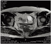
A 30-year-old woman (gravid, 0; para, 0) was referred to us for aggravation of dysmenorrhea during 5 months. Also, she presented with constant dull lower abdominal pain of 5-month duration. She was a virgin and had normal regular menstrual cycles. There was no bowel or urinary symptom. There was no significant medical history, excluding appendectomy 15 years ago. On physical examination, no tenderness was observed. On pelvic examination by rectal, palpable mass with slight tenderness in both adnexa was noted. The transrectal ultrasonography demonstrated a normal uterus, both ovaries and evidenced the presence of round, thick-walled, complex cystic structures measuring 21 × 21 mm, 53 × 34 mm adjacent to the right and left ovaries, respectively. Pelvic computed tomography (CT) and magnetic resonance image (MRI) (Fig. 1) confirmed the aforementioned findings as the pelvic endometriosis.
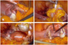
In a view of the history for progressive dysmenorrhea and the psychological impact on the patient, she was counseled and scheduled for diagnostic laparoscopy with the possibility of surgical intervention as deemed necessary. On laparoscopy, the right tube was observed to be twisted twice at its middle part and a thick-walled cystic dilatation at its distal portion that was adherent to the omentum. Symmetrically, the fimbrial end of the left tube was also occluded and three times twisted with congestion as a result, but the uterine isthmic aspect and the midsegment of the tube were identified and were not ischemic (Fig. 2A-2C). The both ovaries with normal appearance were not involved in the torsion and the uterus was normal. Endometriotic implant, such as spot, was only found on the left pelvic side wall. There were no other abnormal findings on laparoscopic abdominal inspection. A laparoscopic bilateral salpingectomy was performed after adhesiolysis (Fig. 2D). The histological examination revealed an extensive hemorrhagic infarction secondary to torsion and the hematosalpinx that endometrial gland was identified.
The postoperative course was uneventful and the patient was discharged home two days later. Until now, she remains well and asymptomatic at follow-up.
Discussion
Isolated fallopian tube torsion is a rare clinical event and the incidence is approximately one in 1.5 million reproductive aged women [2]. Most of the published case reports and occasional series concern unilateral torsion of the isolated fallopian tube. To the best of our knowledge, our case is the first report of isolated torsion of the bilateral fallopian tubes in a reproductive age woman. Our patient had tubal endometriosis which may have obstructed the fimbrial end of the tube and induced in hematosalpinx through retrograde menstruation [4].
The etiology or mechanism of isolated tubal torsion is still uncertain. However, proposed risk factors have been identified. Youssef et al. [5] suggested that intrinsic and extrinsic factors could possibly influence the occurrence of the torsion of isolated fallopian tube. The predisposing intrinsic factors include an excessive length and tortuosity of the tube, hydrosalpinx, hematosalpinx and pyosalpinx, previous sterilization, abnormal peristalsis or endometriosis, while extrinsic factors include paratubal mass, peritubal adhesion, or uterine enlargement compressing the fallopian tubes. Presumably, these factors create a pivot point around which the tube may twist one or several times [6].
The diagnosis of isolated fallopian tube torsion is easily missed preoperatively because of a lack of pathognomonic symptoms, specific findings on physical examination and adequate diagnostic tools. When torsion occurs, the patient usually experiences acute severe lower abdominal or pelvic pain. Pain may be constant or intermittent. Other sings of isolated tubal torsion are inconsistent and comprise anorexia, nausea, vomiting, or vaginal bleeding [7]. Pyrexia, tachycardia, or leukocytosis may be present. Indeed, the clinical presentation often resembles other causes of abdominal pain, for example ectopic pregnancy, pelvic inflammatory disease, ruptured ovarian cyst, hemorrhagic follicle, and acute appendicitis which are far more common. Although transvaginal ultrasound can easily identify enlarged adnexa and Color Doppler can also be used to demonstrate arterial and venous flow to adnexal structure, it must be emphasized that the presence or absence of flow cannot rule out fallopian torsion. CT and MRI are also useful in detecting twisted a vascular pedicle, thickened fallopian tubes, and hemorrhagic infarction. However, the sensitivity of these modalities of the isolated fallopian tube torsion has not yet to be determined [8]. Therefore, most of cases with isolated fallopian tubal torsion had a delayed diagnosis or the diagnosis was not made before surgical intervention because the clinical features are unspecific and objective findings are uncommon. Lo et al. [8] noted that in a total of 17 women with surgically proven isolated fallopian tube torsion, only three women had surgery within 12 hours, but 12 received surgery within seven days; the mean duration of lower abdominal pain until operation was 26.7±58.2 days (range, 0.4 to 180 days). In our case, the time interval between the clinical presentation and postoperative diagnosis of isolated fallopian tube torsion was 5 months.
In the clinical setting of suspected adnexal torsion, emergent laparoscopy is critical to both diagnosis and fertility preservation. Several studies of adnexal torsion have demonstrated that the color, size, and degree of edema do not correlate with necrosis and subsequent return to normal tubal or ovarian function. Immediate detorsion is always recommended, because it is unclear how long a patient has until irreversible damage occurs [6]. However, our patient had bilateral salpingectomy due to obstruction of the both tubal terminal ends and necrosis.
In conclusion, we suggest that in the differential diagnosis of lower abdominal pain in a reproductive age woman, isolated torsion of the fallopian tube should be considered, although it is the low incidence. To the best of our knowledge, our case is the first report of isolated torsion of the bilateral fallopian tubes combined with tubal endometriosis in a reproductive age woman.
 XML Download