

 PDF
PDF ePub
ePub Citation
 Print
Print


Since melanocytes were demonstrated in the cervical epithelium in as many as 3.5% of women [1], malignant melanomas of the cervix were often reported. Approximately 3%-7% of malignant melanomas in women develop within genital tract [2]. The vast majority of these cases occur in vulva and vagina. Cervix is a rare site for melanomas with about 60 cases of malignant melanomas of the cervix have been described as primary tumors within the cervix [3-5]. Malignant melanomas present clinically as an advanced stage and the diagnosis is confirmed by histological examination using special staining and by immunohistochemical study. Radical hysterectomy with regional lymphadenectomy are generally advocated.
Primary malignant melanoma of the cervix has a poor outcome as a consequence of delayed diagnosis and lack of standardized treatment. There is lack of evidence on the efficacy of postoperative radiation or chemotherapy. Radiation therapy may play a role in the treatment of patients with close resection margins, regional nodal metastases or unresectable tumors. Diagnosis is confirmed by immunohistochemical methods and by exclusion of other primary sites of melanomas.
Case Report
A 74-year-old, multiparous woman was admitted to the Department of Obstetrics and Gynecology at Inha University Hospital for vaginal bleeding for several months. A elongated, large sausage like, soft black colored mass with active bleeding and blood clots was noted on the anterior lip of the cervix with suspicious both upper vagina and parametrial invasion. A biopsy from the lesion was taken. The emergency radiation therapy for active cervical bleeding was performed under the impression of cervical squamous cell carcinoma (SCC), stage 1B2, or stage 2B less likely.
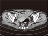
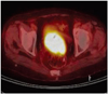
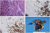
CA-125 and SCC were normal. The abdominopelvic computed tomography (CT) revealed cervical cancer with upper vaginal involvement and suspicious parametrial invasion, and multiple tiny small lymph nodes in both external iliac chain (Fig. 1). Whole body positron emission tomography-CT showed malignancy in the uterine cervix and tiny pulmonary nodule in right medial lobe of lung (Fig. 2). The pathology of cervical punch biopsy showed malignant melanoma. The immunohistochemical staining for S100 protein and HMB-45 antibody were positive, but negative for mixed cytokeratin and leukocyte common antigen (Fig. 3).
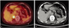
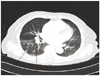
An extensive search for a melanotic lesion including skin and eye was performed to verify the primary site of melanoma, but we could not find any primary site of melanoma. We discussed what is the optimal treatment for this patient with Radio-oncologist and concluded that we would do the surgery because primary melanomas of cervix were poor responders to radiation. We performed the radical hysterectomy with pelvic and paraaortic lymphadenectomy (Fig. 3D). The frozen biopsies of posterior vaginal resection margins were positive tumor involvement in spite of repeated three times of resection in the vaginal cuff. The final pathologic report showed a malignant melanoma, cervix with tumor size 6.0 × 4.7 × 2.0 cm, depth of invasion/thickness of cervix, 2.4 cm/2.5 cm, and tumor involvement of vaginal resection margin, metastatic tumors in 6 out of 38 lymph nodes, positive parametrial, both external iliac and right common iliac lymph nodes. She received postoperative cisplatin based concurrent chemoradiotherapy. Postoperatively 6 months later, her pelvic examination and vaginal stump were clear with good general condition. She had a regular follow-up PET-CT, because there is no tumor markers in malignant melanoma of the uterine cervix. The PET-CT showed metastasis in right adrenal gland and several new pulmonary nodules in right lung (Fig. 4). The chest CT revealed newly appeared multiple well-defined various size pulmonary nodules in right lung and lower lobe of left lung that suspected pulmonary metastasis (Fig. 5). She had a chemotherapy with dacarbazine and cisplatin.
Discussion
Primary melanoma of the cervix is very rare. Melanoma in the uterine cervix may be melanotic or amelanotic [3,6]. About half of the melanomas are amelanotic. Diagnosis of amelanotic melanomas may be difficult due to the absence of pigment.
Patients with cervical melanoma have ranged in age from 19 to 83 years, although the majority have been between 60 to 70 years [3,7,8]. Vaginal bleeding or discharge is the usual presenting complaints. Some patients have been asymptomatic. Patients may remain asymptomatic until the lesions were ulcerated and infected, after then the lesions were easy to bleed. Clinical examination has usually revealed an exophytic polypoid pigmented cervical mass with variable sizes. About half of the cases, brown to blue-black pigmentation of the tumors have been noted. Cervical melanoma is seldom diagnosed by Pap smear in absence of typical pigmented polypoid growth [9]. Morphological features of primary cervical melanoma in Pap smear have been reported, showing bizarre and abnormal cells containing pigment with the hope of early diagnosis [9].
Diagnosis is usually based on pelvic examination and histopathology. Though histological examination in primary cervical melanoma displays predominantly eosinophilic nucleoli, diagnosis should always be confirmed by immunohistochemical staining. A combination of S100 protein (more sensitive) or HMB-45 (more specific) is the best combination for an accurate diagnosis, and is useful in distinguishing amelanotic melanoma from anaplastic carcinoma, high grade lymphoma and sarcoma [7].
Primary cervical melanoma must be differentiated from secondary metastasis of melanoma of other sites in the body, including skin and eye. Norris and Taylor [10] have suggested the diagnostic criterias for primary malignant melanoma: the presence of melanin in the normal cervical epithelium, the absence of malanoma elsewhere in the body, the demonstration of junctional change in the cervix, the metastases according to the pattern of cervical carcinoma.
Histologically the tumors do not differ significantly from melanomas in other sites, being composed of nests of polygonal to spindle-shaped cells, containing mitotically active pleomorphic nuclei. Primary melanoma of the cervix with a prominent spindle cell component should be distinguished from leiomyosarcoma and many benign melanotic lesions, including blue nevus [11]. Primary cervical melanoma has the presence of melanin, junctional activity, immunoreactivity for S100 and HMB-45 antigens, and absence of immunoactivity for markers of smooth muscle differentiation.
There is no consensus on optimal management of the primary malignant melanoma, because of rarity of the lesion. Radical hysterectomy with pelvic and paraaortic lymphadenectomy usually is the most common procedure [12]. There is lack of evidence on the efficacy of postoperative radiation or chemotherapy [13]. Radiotherapy can be used for the palliation of an inoperable patient or as an adjuvant therapy [14]. Dacarbazine which has been shown to reduce tumor in patients with cutaneous malignant melanoma may be useful for cervical malignant melanoma [4,12]. A combination chemotherapy with cisplatin, vinblastin and bleomycin may produce a better response than a single agent dacarbazine [3,12]. The prognosis of the primary cervical melanoma is generally poor, because diagnosis is usually made at advanced stage and the tumor is highly aggressive as both local recurrence and widespread metastases [6]. The 5-year survival rate after radical hysterectomy is not exceeding 40% in stage 1 and reaching only 14% in stage 2 [7]. The reported survival time ranges from 6 months to 14 years, 90% of the reported patients with follow-up data have been died of their diseases, within 2-3 years of presentation [12].
In conclusion, primary malignant melanoma of the cervix should be considered in the differential diagnosis of cervical malignancies. The diagnosis should be confirmed using special stains and immunohistochemistry. This is essential since cervical melanoma is incurable even with the currently available therapies and hence needs to be diagnosed early.
 XML Download