

 PDF
PDF ePub
ePub Citation
 Print
Print


Ectopic pregnancy refers to the implantation of a viable embryo outside the uterine corpus. Heterotopic pregnancy is diagnosed in the presence of simultaneous gestations at two or more implantation sites. Its occurrence is rare in spontaneous conception with an incidence of 1:30,000, while the incidence is found to be as high as 1% in pregnancy by assisted reproductive technology [1].
We report a case of a spontaneous heterotopic pregnancy that was diagnosed by visualization of heart activity in both intrauterine and extra-uterine gestations.
Case Report
A 34-year-old woman gravida 1, para 1, was admitted to our emergency center with a complaint of severe lower abdominal pain, vaginal spotting, nausea and vomiting. She had irregular menstrual cycles and was amenorrhea for 8 weeks after last menstrual period. She did not check urine pregnancy test before visiting hospital. She gave a full-term vaginal birth without complications and she had an ectopic pregnancy which was treated with methotrexate two years before. She desired a baby and therefore had never been on birth control. The patient had been in a long-term healthy, monogamous relationship and denied any history of sexually transmitted diseases and pelvic inflammatory disease. She denied any medical problems, previous surgeries and use of tobacco, alcohol, or illicit drugs. Her family history was unremarkable for any pelvic diseases.
On examination, she was pale with a pulse rate 100/min, blood pressure 100/60 mm Hg, respiratory rate 24/min and body temperature 37.2℃. Physical examination revealed diffuse lower abdominal tenderness with signs of peritoneal irritation. Enlarged, tender uterus corresponding to 8 weeks of pregnancy was detected in the pelvic examination. In addition, a tender mass was also palpable in her right adnexa. Cervical movement during the bimanual examination caused severe pain. On speculum examination there was minimal darkish bleeding from the cervical os.
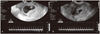
Transvaginal ultrasound showed the intrauterine embryo with cardiac activity corresponding to 8 weeks of gestation by crownrump length (Fig. 1A) and an extra-uterine right adnexal embryo with cardiac activity corresponding to 7 weeks of gestation (Fig. 1B). Also, accumulation of fluid was noted in the Pouch of Douglas. Her hemoglobin was 9.1 g/dL with a normal white blood cell count and platelet count. Serum β-human chorionic gonadotropin (hCG) was 17,036 mIU/mL.
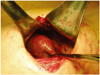
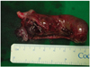
She underwent emergency exploratory laparotomy under the diagnosis of heterotopic pregnancy. During surgery, a right abortive tubal ectopic pregnancy with hemoperitoneum was identified (Fig. 2) showing active bleeding in the ampulla portion. Intra-abdominal bleeding was so heavy that it was deemed dangerous to preserve the tube and right salpingectomy was carried out. After hemostasis and irrigation, the abdomen was closed. The histologic examination of the salpingectomy confirmed the diagnosis (Fig. 3).
The postoperative course was uneventful. On the 2nd postoperative day, transvaginal ultrasound confirmed heart activity of a living intrauterine pregnancy. She was discharged on the 5th postoperative day without problem in stable condition. Two weeks after surgery, transvaginal ultrasound revealed continued normal growth of the intrauterine embryo, but she was lost in follow-up after then.
Discussion
The ectopic pregnancy can be tubal, ovarian, cervical, cornual, or abdominal. About 1% of the pregnancies are in an ectopic location, of which 95%-97% are located in the fallopian tube. The most common site is the ampulla portion of the tube (80%), followed by the isthmic segment of the tube (10%), the fimbria (5%) and the cornual and interstitial regions (2%-4%) [2]. Common factors that predispose to occurrence of ectopic pregnancy are tubal surgery and pelvic inflammatory diseases.
The heterotopic pregnancy is a combination of an intra-uterine and extra-uterine pregnancy, at the same time. The spontaneous heterotopic pregnancy is a rare illness with an estimated frequency below one per 20,000 and one per 30,000 [3]. The first case was reported in France by Duverney in 1708 during an autopsy [4]. Although spontaneous simultaneous intrauterine and ectopic pregnancy was an extremely rare event in the past, it is increasingly being diagnosed since the rate of assisted reproductive technique increased. As in our case, when previous ectopic pregnancy has been treated with methotrexate, future pregnancy is associated with an increased risk for ectopic pregnancy and potentially heterotopic pregnancy [5].
The preoperative diagnosis of a heterotopic pregnancy remains a major challenge for modern reproductive medicine. Although signs and symptoms such as abdominal pain, adnexal mass, peritoneal irritation, vaginal bleeding, and enlarged uterus have been reported to be predictive of a heterotopic pregnancy, they are nonspecific and may be confused with other normal or abnormal pregnancy manifestations. Furthermore, if assisted reproductive technique is not involved, the index of suspicion of multiple or heterotopic pregnancy is usually very low. Heterotopic pregnancy is a rare condition and most patients present in the emergency department with symptoms of a rupture of ectopic component [6]. Thus, a preoperative diagnosis of heterotopic pregnancy is still a challenge.
The advent of ultrasound for pregnancy positive patient has not changed diagnostic ability over a period of time. In a review of the literature of all cases of heterotopic pregnancy from 1971 to 1993, out of 112 cases, 46 were diagnosed by ultrasound while 66 were diagnosed at laparoscoy or laparotomy [7]. Transvaginal ultrasound and assesment of the whole pelvis, even in the presence of intrauterine, can be an important aid in the diagnosis of heterotopic pregnancy. Visualization of heart activity in both intrauterine and extra-uterine gestation, as in our case, confirms the diagnosis of heterotopic pregnancy. Serial β-hCG levels are not of much significance and diagnosis of heterotopic pregnancy as subnormal hormone production by an ectopic pregnancy may be masked by the higher placental production from the intrauterine pregnancy [8]. Culdocentesis is an important aid in diagnosis when hemoperitoneum is presented as echogenic cul de sac fluid collection is more important than anechoic fluid because it indicates the presence of peritoneal hemorrhage.
Management of heterotopic pregnancy still remains controversial. The standard treatment for ectopic pregnancy is surgery by laparoscopy or laparotomy depending upon the condition of the patient. After diagnosis, the ectopic component is usually treated surgically, whereas the intrauterine pregnancy is expected to develop normally. The main aim of the surgery should be the preservation of the intrauterine pregnancy with minimal manipulation of the uterus [9]. Many cases are treated by surgery via laparoscopy or laparotomy, including salpingotomy or salpingectomy. The choice between conservative or radical treatments may be difficult. However, a recent review demonstrated no difference in rates of intrauterine pregnancies after conservative or radical surgery for tubal ectopic pregnancy [10]. It seems that, particularly in patient with an intact contralateral tube, fertility results after salpingectomy are comparable to those observed after salpingotomy [11]. Moreover, radical treatment is easier, thus reducing the risk of complication observed at salpingotomy. Laparotomy has been used widely until recently. Nowadays, laparoscopy is preferred treatment for heterotopic pregnancy and laparoscopy is the appropriate modality both for diagnosis and treatment of heterotopic pregnancy [12]. However, laparotomy may be the preferable surgical modality in cases with serious intra-abdominal bleeding or in patients with hemorrhagic shock [13]. In the present case, we preferred laparotomy because of the presence of signs serious intra-abdominal bleeding. In conclusion, heterotopic pregnancy should be thought in differential diagnosis of an acute abdomen. It is important to remember that the detection of an intra-uterine pregnancy does not exclude the existence of an accompanying ectopic pregnancy. In the case of heterotopic pregnancy, salpingectomy should be considered when the contralateral fallopian tube is healthy as this treatment does not preclude future fertility.
 XML Download