

 PDF
PDF ePub
ePub Citation
 Print
Print


Vulvar varices are reported in 4% of women [1], and most of them may be secondary to pregnancy from other anatomical and pathological diseases such as Klippel-Trenaunay-Weber syndrome or pelvic congestion syndrome. However, the vaginal varices, located in the vaginal and periurethral wall, are reported less frequently. Vaginal varix in non-pregnant women does not exhibit a serious threat; however, during pregnancy, the spontaneous laceration of the vaginal varix can cause significant blood loss. Therefore, an obstetrician should be aware of its clinical importance. We report a case of vaginal varix during pregnancy and its clinical features for the first time in Korea.
Case Report
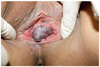
A 32-year-old multiparous woman who was a nonsmoker with no significant medical history visited the department of obstetrics and gynecology because of acute spontaneous vaginal bleeding. The woman was at 37+6 week of gestation with the fetus having a cephalic presentation. The pregnancy had been normal during the prenatal check 2 weeks ago, when the patient experienced cough and rhinorrhea. The patient recognized the lesion had been enlarged as the pregnancy continued few months into the conception. We found the vaginal bleeding and performed a pelvic inspection by spreading the labia minor with two fingers, and were surprised to find a strawberry sized vascular lesion on the suburethral vaginal wall, obstructing the vaginal lumen (Fig. 1). We detected a bleeding point on the varix.
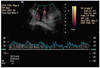
On the visit, the patient's vital signs were stable and the fetal heart rate pattern on non-stress test was reactive. The sonographic examination revealed a mixed echogenic mass that was 2.3 × 2.7 cm in size at the anterior vaginal wall area. Maximal systolic and diastolic velocity at 21.7 cm/sec and 12.8 cm/sec, respectively, was found using Power Doppler sonography, and the resistance index was 0.41 (Fig. 2). The fetus grew normally according to the gestational age. Also, the placenta showed left lateral attachment and the amniotic fluid index was measured as 16.78 cm. We sought the opinions of a gastroenterologist and a vascular surgeon, who both agreed on the diagnosis of vaginal varix.
The prevalence of varices on the esophagus and the legs led us to perform additional tests, such as the gastrofundoscopy to seek any esophageal lesion, with negative finding. A Doppler ultrasound on both the lower extremities was done to rule out any other varices at the lower limbs. There was no visible venous reflux or perforating vein in both great saphenous veins.
We then performed a Cesarean section with a low transverse incision to avoid intrapartum bleeding, and did not run into any other problems. Following the delivery, a routine dose of uterotonics was administered. The vaginal varix did not shrink immediately after the cesarean section. The patient wore elastic stockings for the remainder of the stay to prevent any thromboembolic disease. At 6 weeks postpartum evaluation, the speculum examination of the vagina showed the disappearance of the vaginal varix.
DISCUSSION
Pregnancy is associated with dilatation of the vascular system as a result of hormonal influences and increased blood volume. Varices are often exacerbated during pregnancy, because the enlarged uterus compresses the pelvic veins and the inferior vena cava [2].
The occurrence of the varices at the vaginal wall is rare, although it is more common on the vulvar area. Only a few cases have been reported in the literature, and these cases exhibited vaginal bleeding from the varices, as a complication of portal hypertension [3,4]. In contrast, our patient displayed neither portal hypertension nor any other varices.
Genital varices usually appear during the third or fourth month of gestation and regress spontaneously after delivery [1]. In this case, the patient recognized the lesion growing in the later months of gestation. Most genital varices are asymptomatic, but a few are associated with severe local discomfort, accompanied by spontaneous vaginal bleeding, or with pelvic pain as part of the so-called "pelvic congestion syndrome"; pelvic pain, dyspareuria, dysmenorrhea, dysuria, vulvar and perivulvar varices [5,6].
The vaginal varix can be ruptured from trauma, leading to a vaginal hematoma or thrombus during the second stage of labor. This hematoma or a thrombus can result in a subsequent extravasation into the tissues. The bleeding from the vaginal varices may have a profound feto-maternal effect if the veins are large and the walls are thin. Another case was reported with a fetal death from significant maternal bleeding from the varix rupture, with the bleeding spot seen as a hole in a localized varicosity of a vein [7]. Watermeyer et al. [8] reported a 21-year-old primiparous woman with a massive plexus of varicosities filling the uterovesical fold that extended over the lower uterine segment and performed a classical Cesarean section with a blood loss of 1,500 mL. This patient experienced spontaneous vaginal bleeding after coughing infrequently. The patient did not suffer from excessive blood loss.
It is important to recognize the possibility of associated anatomical or pathological diseases. It may include leg varices, venous malformation of the labia, clitorial area, or vagina with or without arteriovenous malformation on the limbs or trunk (Klippel-Trenaunay syndrome) [8]. The Klippel-Trenaunay syndrome consists of a triad of cutaneous port-wine capillary malformations, varicose veins and hemihypertrophy of soft tissue and bone. The diagnosis can be made if two of the above three features are found.
The lesion should be compared against a few possible options, such as a hemangioma or angiosarcoma. One of the diagnostic methods is to take a biopsy of the lesion. However, the risk of causing vaginal bleeding compelled us to attempt a less invasive way to make a diagnosis [9]. Thus, the Doppler study was considered. A varix is of the venous family, thus would therefore not demonstrate any pulse. However, it was interesting to see such a difference in magnitude. We postulate that the noise from the surrounding small arteries clouded the data. In the current state, even though a duplex ultrasound is useful in the diagnosis of the varix, a history and physical examination findings are still important. Therefore, the authors reiterate the diagnosis of varix on this patient from physical examination.
The treatment of the vaginal and vulvar varix is generally conservative; firm support and application of pressure may be enough to relieve the symptoms. If the symptoms persist for more than 12 weeks postpartum, the varix can be treated with sclerotherapy [10]. Although in some mild cases successful treatment can be achieved with either local excision or sclerotherapy [5,6,10,11], a laparoscopic ligation of the incompetent veins can be considered in patients with pelvic congestion syndrome [11]. In the author's case, the patient's vaginal varix resolved after 6 weeks postpartum without any additional treatment.
Obstetricians need to be aware that the possibility of vaginal varix can exist in patients complaining of vaginal bleeding and that vulvar varicose veins can be a part of the Kippel-Trenaunay syndrome which require long-term anticoagulation and some specific elastic stockings to prevent thromboembolic events after delivery. We realize the need for a less invasive and more accurate way to confirm vaginal varix. Therefore, we report this case with a brief review on vaginal varices.
 XML Download