

 PDF
PDF ePub
ePub Citation
 Print
Print


Hydrometrocolpos is a condition in which the uterus and vagina are distended by retained fluid other than blood in the presence of distal vaginal outlet obstruction. Secondary infection of hydrometrocolpos leads to pyometrocolpos (pyometra). The condition is rarely diagnosed in the neonate or infant until the obstruction causes a significant fluid collection in the vagina above the obstructing septum and cause compression effect to the surrounding structures. Diagnosis is made initially by abdominal ultrasonography but magnetic resonance imaging (MRI) is used for definitive diagnosis. It can be diagnosed prenatally, during the third trimester by transabdominal sonography [1]. In children the hydrometrocolpos present usually as suprapubic cystic mass with or without retention of urine which may be an acute emergency [2]. Urgent drainage of the infected cystic mass leads to marked improvement in the clinical course of such patients who require definitive surgery at a later stage [3]. Rarity and variable presentations can lead to delayed diagnosis and erroneous management.
Case Report
A 2-year-old girl who reported to the paediatrics outpatient department (OPD), was referred to the Department of Obstetrics and Gynaecology with complaints of a mass in suprapubic region and acute retention of urine for two days. Intermittent catheterisation was done by plain rubber catheter before her examination in the OPD. Physical examination of the patient revealed age-appropriate developmental milestones. Patient was severely pale and febrile. Her pulse rate was 110 beats per minute, regular and temperature was 100 degree F. Her cardiovascular and respiratory examination was normal except for a pan systolic murmur. On per abdominal examination a suprapubic tender mass was felt.
On local examination, her external genitalia appeared normal. All her routine laboratory investigations were within normal limits except for a haemoglobin level of 5.7 g/dL, total leucocyte count was 5,700/mm3, Differential leucocyte count was P60L37E03M00 and C-reactive proteins was positive. Urine routine and microscopic examination was normal and culture and sensitivity was sterile.
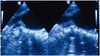
Ultrasonography (USG) of the whole abdomen and pelvis showed a distended uterine cavity suggestive of haematometrocolpos with absent left kidney (Fig. 1). No other anomaly was noted. MRI pelvis and computed tomography (CT) was advised but patient was very poor so refused for above investigations. Urgent drainage of the haematometrocolpos was planned. The patient was shifted to operation theatre where transurethral catheterisation was done.
Pelvic examination under general anaesthesia showed a low transverse vaginal septum. On per rectal examination a soft cystic mass was felt in the suprapubic region. A 10 mL syringe was introduced through the transverse vaginal septum and pus was drained which was sent for bacterial and acid fast bacillus culture and sensitivity. To rule out tubercular pyometra acid fast bascilli (AFB) culture and sensitivity of pus was done which turned out to be negative.
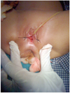
The transverse vaginal septum was excised and about 100 mL pus was drained (Fig. 2). Upper and lower vaginal mucosa was anastomosed. Triple antibiotic coverage was given and the patient received two units of blood transfusions post operatively. Pus culture showed the growth of Eischerecia coli which was sensitive to amikacin as well as ceftriaxone. For anaerobic coverage metrogyl was also added. Marked improvement in the general condition of the patient was seen in the immediate post operative period. Silicone vaginal form was applied and patient's mother was educated for vaginal form. The girl was discharged in satisfactory condition with advice for follow-up but she did not turn up.
Discussion
Developmental anomalies of the mullerian duct system represent some of the most fascinating disorders that gynaecologists encounter. Mullerian malformations are frequently associated with abnormalities of the renal and axial skeletal system and are often first encountered when patients are initially examined for associated conditions. Disorders of vertical (transverse) fusion result from abnormal canalization of the vaginal plate and, in some cases, failure of the uterovaginal primordium and the sinovaginal bulbs to fuse. These disruptions can result in the formation of a transverse vaginal septum (TVS), an imperforate hymen, and in extreme cases, vaginal atresia. The TVS can occur at nearly all levels in vagina, the most common location being superior vagina (46%), then mid vagina (40%), and the most rare is inferior vagina (14%) [1].
Congenital obstructing lesions of the vagina lead to accumulation of fluid distending the vagina which is known as hydrocolpos or mucocolpos. If the volume of secretions is so large that the uterus is also distended, this condition is called hydrometrocolpos. Secondary infection of hydrometrocolpos leads to pyometrocolpos which can present as an emergency [2,3] as happened in our case. Hydrometrocolpos and haematocolpos caused by imperforate hymen or distal vaginal atresia present with recurrent urinary tract infection, abdominal mass and sometimes findings of obstructive uropathy during the neonatal period as seen in our case but the condition often go unrecognised until weeks or months later when they develop the above three hallmarks of the condition. True incidence of these anomalies is unknown but is reported to be between 0.1% and 3.8% [4]. The incidence of hydrometrocolpos is 1 in 16,000 live births [5]. The rarity of this anomaly quoted in the earlier literature was probably due to difficulty in diagnosis and high mortality rates resulting from infections and associated anomalies even before the diagnosis could be established.
Complication of pyometrocolpos requiring urgent attention among both infants and adolescents is acute urinary retention. Rarely, an infant may have respiratory insufficiency or inferior vena cava compression because of the large mass. Endometriosis can be a late complication due to retrograde blood flow especially in patients with high transverse vaginal septum [1]. Diagnosis can be challenging. Initial studies should include a USG of the abdomen and pelvis. MRI can also be useful to depict pelvic anatomy and determine the thickness of the vaginal septum to arrive at a definitive diagnosis [6]. CT scan is useful to demonstrate the cystic mass distending the uterus and vagina. Colour Doppler rules out associated endometrial malignancy. Intravenous pyelography to be done for associated abnormalities of the renal system. To rule out tubercular pyometra AFB culture and sensitivity of pus should be done.
Patients with congenital vaginal obstruction need surgical treatment which depends on its location and thickness. In infants and neonates the TVS is usually thin and can be excised without extensive procedure through perineal approach under local anaesthesia. The TVS can be thick in adults and require more extensive approach depending upon its location in the vagina. High haematocolpos which can not be drained vaginally will require exploratory laparotomy under general anesthesia. While mid and low TVS are excised with multiple radial incisions with anastomosis of upper and lower vaginal mucosa. In this case the transverse vaginal septum was thin which was excised and about 100 mL pus was drained. Upper and lower vaginal mucosa was anastomosed and silicone vaginal form was applied. Patient's mother was educated for vaginal form. As the girl did not turn up for follow up we could not see the status of vagina after the constrictive phase is over. Immediate drainage of the cystic mass in a patient with pyometrocolpos is required to prevent or treat uropathy and septicaemia until definitive corrective surgery can be performed. Vaginal reconstruction may be required to allow satisfactory menstruation and coitus.
 XML Download