

 PDF
PDF ePub
ePub Citation
 Print
Print


Schwannoma, neurinoma or neurilemmoma arise from schwann cells in the peripheral nerve sheath, without involving the nerve fibers.
Its most frequent sites are the extremities, posterior mediastinum, retroperitoneum, head and neck. On the other hand, schwannoma is a benign tumor which rarely affects the female genitalia [1,2]. Especially, despite the rich nerve supply of the clitoris, schwannomas of the clitoris are extremely rare [3-5]. We report a case of schwannoma of the clitoris.
Case Report
A 76-year-old postmenopausal Korean woman, who had four children, was referred to our hospital in 9 February 2010 for evaluation of vaginal spotting.
Physical examination revealed a 2.0 × 1.7 cm sized, movable, non-tender mass in the clitoris. But, she had not noticed the clitoral mass before; it was not painful.
Except for this lesion, her external genitalia was normal. She underwent hysterectomy 15 years before due to uterine prolapse. Her vagina was clear. There was no definite bleeding focus. Also, no abnormalities were detected on neurologic examination. She had no urinary or gastrointestinal problems. And there was no evidence of virilism and neurofibromatosis. About 7 years ago, she was diagnosed with myocardial infarction, and she has been taking heart medicines.
There was no family history of virilism or neurofibromatosis. And there was no family history of other hereditary disease. But, her brother died of lung cancer.
On 9 February 2010, simple surgical excision of the clitoral mass was performed under local anesthesia.
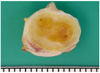
The specimen was a well demarcated, encapsulated and yellowish myxoid nodule measuring 2.0 × 1.7 × 1.5 cm. There was no hemorrhage or necrosis (Fig. 1).
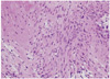
Microscopically, features of schwannoma were observed. The tumor was composed of many spindle cells with nuclear palisading arranged in whorls (Fig. 2).
There was no postoperative complication. And no evidence of local recurrence was noted after 1 year of follow-up.
DISCUSSION
Schwannoma, also known as neurinoma, neurilemmoma, neurolemmoma [6] and perineural fibroblastoma [7] is a peripheral nerve sheath tumor characterised by proliferation of schwann cells [7-10].
Schwannomas rarely affect the female genitalia [1,2]. Especially, despite the rich nerve supply of the clitoris, schwannomas of the clitoris are extremely rare [3-5].
Schwannomas are benign, slow-growing tumors that infrequently recur and rarely undergo malignant change [11].
Histologically, the distinctive feature of schwannoma is the pattern of alternating Antoni A and B areas [7-10]. Antoni A areas are composed of compacted spindle cells arranged in palisades or in an organoid arrangement (verocay bodies). Antoni B areas consist of tumor cells suspended in a loosely textured matrix with a more rounded cell morphology. Our case also showed that the tumor was composed of many spindle cells with nuclear palisading arranged in whorls.
Also, an immunohistochemical examination can be used in the diagnosis. In addition to the S-100 protein, positivity tumor cells express basal lamina components (such as laminin and type IV collagen), vimentins, and sometimes KP1 (CD68) and glial fibrillary acidic proteins [5].
The most common types of benign peripheral nerve sheath tumors are schwannoma and neurofibroma [7,8]. But microscopically, the presence of encapsulation, two types of Antoni areas, and diffusely strong immunostaining for S-100 protein distinguish schwannomas from neurofibromas [12]. And clinically, most of the clitoral neurofibromas occur in a background of neurofibromatosis, mostly neurofibromatosis type 1 [13], whereas none of the reported cases of clitoral schwannoma had neurofibromatosis. Neither in this case. In conclusion, despite its rarity, a schwannoma should be included in the differential diagnosis of a clitoral mass [14]. And simple surgical excision and follow-up considered as most adequate treatment [3].
 XML Download