

 PDF
PDF ePub
ePub Citation
 Print
Print


Embryonal Rhabdomyosarcoma (RMS) is the most common soft tissue tumor in childhood and young adults. RMS is rare in adults, with soft-tissue sarcomas making up less than 1% of malignancies in adults and RMS accounting for 3% of all soft-tissue sarcomas. In University of Tehran data only 6 RMS were found (0.39%) among the 1,528 patients with genital tract malignancies [1]. In Korea, a total of 8 cases of RMS were reported in young adults but there has been no case in which the patient was older than 40 years.
We report a case of cervical RMS in a 52-year-old woman who underwent radical surgery.
Case Report
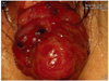
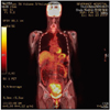
A 52-year-old multiparous woman presented with vaginal bleeding for several weeks and the feeling of a mass protruding from the introitus for 2 months. She has normal menstrual cycles. The patient was referred to our institute with a suspicion of a cervical myoma. In her medical history, she had been diagnosed with cerebral arteriovenous malformation and treated with embolization and gamma knife surgery a few months prior. On the vaginal examination, a 7 × 6 cm soft pinkish irregular mass arising from the uterine cervix and reaching up to the introitus was seen (Fig. 1). Biopsy was performed and the result was favoring sarcoma, most likely embryonal RMS. Magnetic resonance imaging and F-18 flurodeoxyglucose (FDG) positron emission tomography/computed tomography also showed a 7 × 6 cm mass arising from the endocervix and protruding outside the vagina cavity to the perineal portion with FDG uptake (Fig. 2). Moreover, parametrial invasion was seen on posterior aspect of the uterine cervix. The serum level of CA-125 was 58.6 U/mL. Other laboratory tests showed no specific abnormality. Her height, weight, and body mass index were 156 cm, 53 kg, and 21.8 kg/m2, respectively. Based on the results of these imaging studies and the pathologic result of biopsy, the patient was diagnosed with sarcoma of the uterine cervix. Therefore, radical abdominal hysterectomywith bilateral salpingo-oophorectomy, and bilateral pelvic lymph node dissection were performed. Additionally, radical vaginectomy wasperformed to ensure adequate resection margin. The final pathologic result was cervical RMS, consistent with the Intergroup RMS Study Group IIC. Immunohistochemistry was positive for desmin, myogenin andmyogenic diffentiation 1 (myoD1). Concurrent chemoradiation therapy was planned. The regimens for chemotherapy was vincristine of 1.5 mg/m2 on the first day plus dactinomycin of 0.3 mg/m2 and cyclophosphamide of 150 mg/m2 from first day until fifth day for 6 cycles. However, she has been observed without adjuvant treatment due to cerebral hemorrhage and relapsed septic condition after surgery. She received conservative management in a convalescent hospital.
1. Histopathologic results
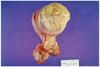
There was a grossly huge pedunculated mass with surface nodularities, measuring 7 × 5.5 × 5 cm, arising closer to endocervix than the junction of the lower uterine segment (Fig. 3). On sections, it was pinkish gray myxoid and showed a multifocal hemorrhage. A well-demarcated bulging intramural mass, measuring 11 × 2 cm was noted in the myometrium. Microscopically, the intramural leiomyoma was involved by the RMS. The desmin immunoreactivity confirmed the myoblastic differentiation. The tumor consisted of oval to spindle shaped malignant embryonal rhabdomyoblasts with eosinophilic cytoplasm (Fig. 4A). Higher magnification of the tumor cells showed cross striation. Histopathological picture favoured embryonal RMS, The soft tissue of right ureteral, left uterosacral, parametrium, anterior and posterior vaginal wall, vaginal cuff, and right uterine artery were also infiltrated by tumor. The lymphovascular space invasion was noted. Pelvic lymph nodes were free of tumor. Immunohistochemistry was positive for phosphotungstic acid-hematoxilin, myoD1, myogenin, desmin, and smooth muscle actin (Fig. 4B).
Discussion
The genitourinary tract is the second most common site of RMS. The peak incidence age of cervical RMS is the second decade and uterine corpus RMS is most common at menopause phase.
The corresponding tumor in the vagina usually occurs in infants and young children and establishing the diagnosis may be difficult in an older age group without clinical suspicion of malignancy and in whom cervical polyps are common. The initial clue to the diagnosis is often the gross appearance, in that the polyp may be unusually large and myxoid or there may be multiple polyps. Clinical manifestations of RMS are serosanguinous vaginal discharge and polyp-like mass protruding through vagina. In advanced disease, pain, bladder irritation, and tenesmus were noted. In terminal status, hydronephrosis, anemia, and cachexia were developed.
Survival rate of RMS depends on the site of origin, size, extent of disease, residual disease after treatment, histologic subtype and anaplasia. In vaginal lesions, a better prognosis has been documented than in cervical lesions [2].
The most widely used classification for RMS is that of Horn and Enterline which consists of four histologic subtypes: embryonal, botryoid subtype of embryonal, alveolar, and pleomorphic RMS [3]. There are distinct histological subtypes of RMS that differ in their clinical presentation and behavior. Overall, the embryonal subtype is more common in young children accounting for up to 49% of all RMS [4] and has a better prognosis. The alveolar RMS is secondary most common and more common in adolescents that incidence is about 25% and that is frequently metastatic at diagnosis, and has a worse prognosis. The pleomorphic subtype predominates in adults, among whom RMS is relatively infrequent [5].
According to Ghavimi et al. [5] all patients were treated according to protocols consisting of surgery, radiotherapy, and multiple drug chemotherapy, overall survival rate was 63%. Early stage embryonal RMS of the cervix has been found to have an excellent prognosis. Zeisler et al. [6] demonstrated that the survival rate was 92% in early stage RMS after local excision with chemotherapy. The estimated 5-year survival rates for patients with and without anaplasia were 68% [7].
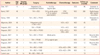
Only eleven cases of uterine cervix RMS in women over age of 40 years have been reported (Table 1) [8]. We present here the 12th case. As the patient had extensive disease we opted to perform a radical hysterectomy with pelvic lymph node dissection although its role has no consistent data in literature. Our case is the third patient over 40 years of age who was treated with radical hysterectomy followed by the Intergroup Rhabdomyosarcoma Study group IV adjuvant chemotherapy schema [9].
Recently in Korea, Jang et al. [10] reported a RMS in 2001 but it was a case in a 31 year-old woman. In the present case, the patient was a middle-age woman who underwent radical surgery without adjuvant treatment. This is an unusual case of RMS and the prognosis of the patient needs to be carefully monitored on follow-up.

 XML Download