

 PDF
PDF ePub
ePub Citation
 Print
Print


Abstract
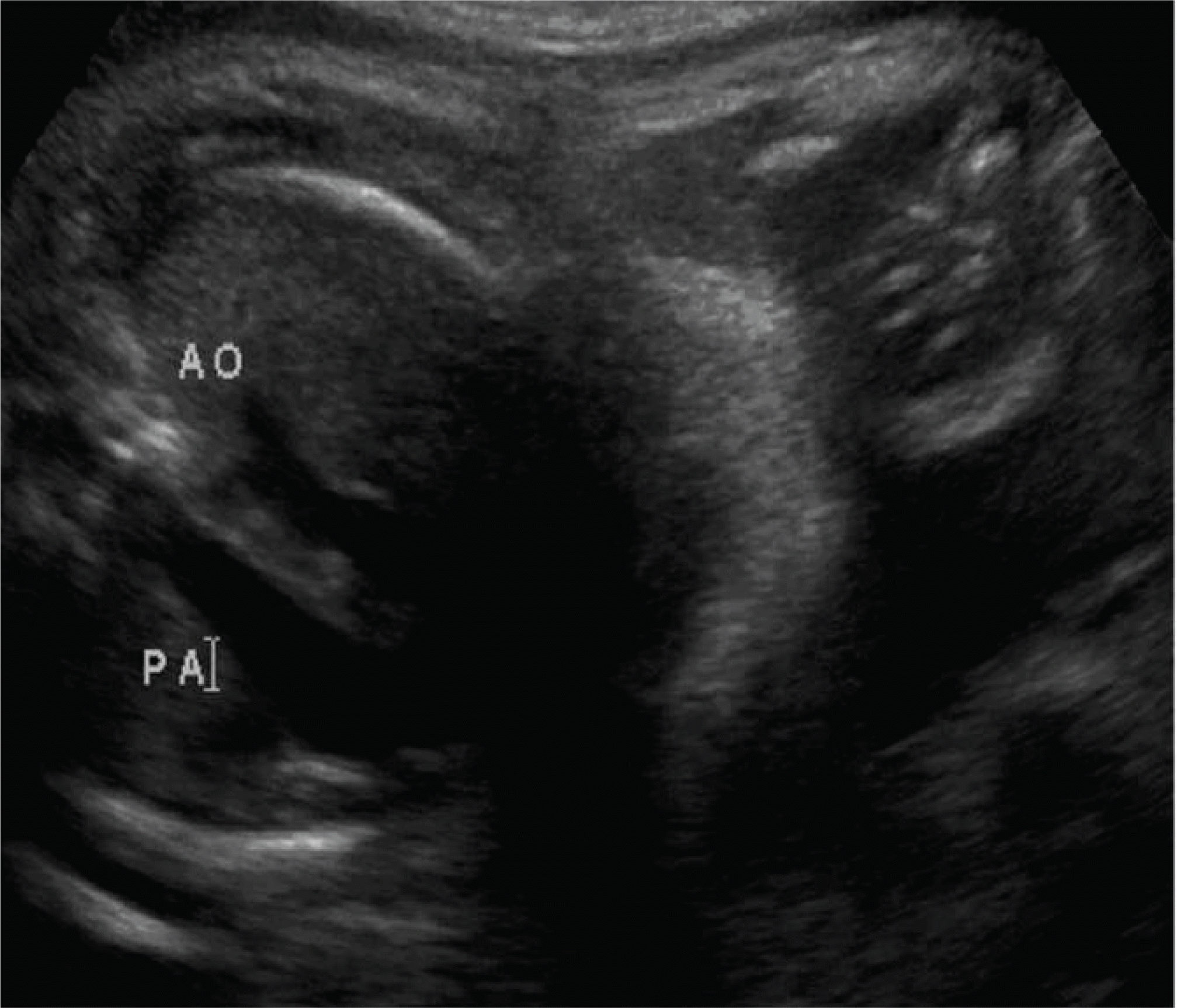
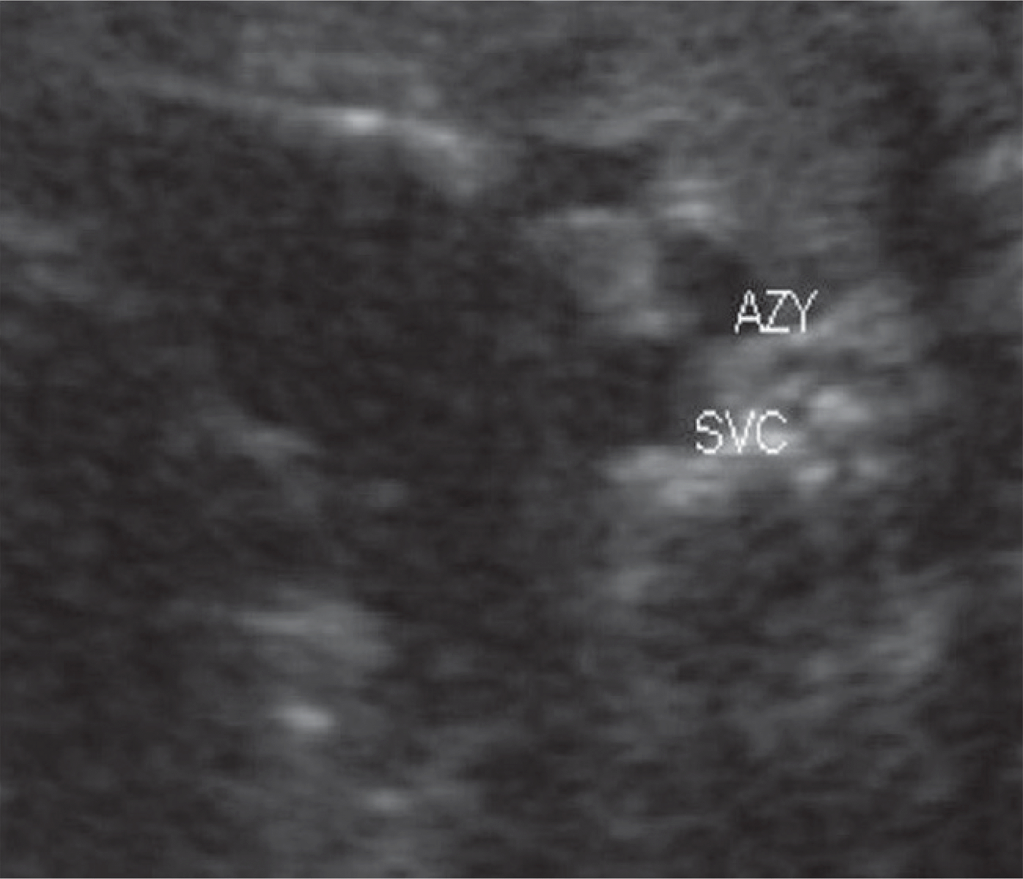
Heterotaxy syndrome is a disorder that results in abnormal placement of organs. We report the prenatal diagnosis of complex cardiac abnormalities associated with left isomerism. A 32-year-old multigravida was referred to our hospital for evaluation of abnormal sonographic findings. On prenatal sonography, we suspected a double outlet of the right ventricle, right ventricular hypertrophy, a ventricular septal defect, a large azygos vein instead of an inferior vena cava, a right-sided stomach, and asplenia. Postnatal imaging studies confirmed these findings, except a left-sided single spleen. After birth, the infant underwent a left modified Blalock-Taussing shunt and underwent a 2nd surgical procedure (Kawashima operation, right pulmonary artery angioplasty, and atrial septal defect widening). This report suggests intrauterine fetal sonography can accurately delineate abnormal findings in heterotaxy, allowing perinatal counseling and postpartum planning for corrective surgery.
REFERENCES
1. Bowers PN, Brueckner M, Yost HJ. The genetics of left-right development and heterotaxia. Semin Perinatol. 1996; 20:577–88.

2. Lin AE, Ticho BS, Houde K, Westgate MN, Holmes LB. Heterotaxy: associated conditions and hospital-based prevalence in newborns. Genet Med. 2000; 2:157–72.
3. Berg C, Geipel A, Smrcek J, Krapp M, Germer U, Kohl T, et al. Prenatal diagnosis of cardiosplenic syndromes: a 10-year experience. Ultrasound Obstet Gynecol. 2003; 22:451–9.
4. Cohen MS, Anderson RH, Cohen MI, Atz AM, Fogel M, Gruber PJ, et al. Controversies, genetics, diagnostic assessment, and outcomes relating to the heterotaxy syndrome. Cardiol Young. 2007; 17(Suppl 2):29–43.
5. Taketazu M, Lougheed J, Yoo SJ, Lim JS, Hornberger LK. Spectrum of cardiovascular disease, accuracy of diagnosis, and outcome in fetal heterotaxy syndrome. Am J Cardiol. 2006; 97:720–4.
6. Tongsong T, Sittiwangkul R, Wanapirak C, Sirichotiyakul S. Prenatal diagnosis of transposition-like double-outlet right ventricle with mitral valve atresia in heterotaxy syndrome. J Clin Ultrasound. 2005; 33:197–200.
7. Lim JS, McCrindle BW, Smallhorn JF, Golding F, Caldarone CA, Taketazu M, et al. Clinical features, management, and outcome of children with fetal and postnatal diagnoses of isomerism syndromes. Circulation. 2005; 112:2454–61.
8. Stumpflen I, Stumpflen A, Wimmer M, Bernaschek G. Effect of detailed fetal echocardiography as part of routine prenatal ultrasonographic screening on detection of congenital heart disease. Lancet. 1996; 348:854–7.
9. Buskens E, Grobbee DE, Frohn-Mulder IM, Stewart PA, Jutt-mann RE, Wladimiroff JW, et al. Efficacy of routine fetal ultrasound screening for congenital heart disease in normal pregnancy. Circulation. 1996; 94:67–72.
10. Cohen MS. Clarifying anatomical complexity: diagnosing heterotaxy syndrome in the fetus. Prog Pediatr Cardiol. 2006; 22:61–70.
11. Yoo SJ, Jaeggi E. Ultrasound evaluation of the fetal heart. Callen PW, editor. Ultrasonography in obstetetrics and gynecology. 5th ed.Philadelphia (PA): Saunders Elsevier;2008. p. 523–8.
12. Montaña E, Khoury MJ, Cragan JD, Sharma S, Dhar P, Fyfe D. Trends and outcomes after prenatal diagnosis of congenital cardiac malformations by fetal echocardiography in a well defined birth population, Atlanta, Georgia, 1990-1994. J Am Coll Cardiol. 1996; 28:1805–9.
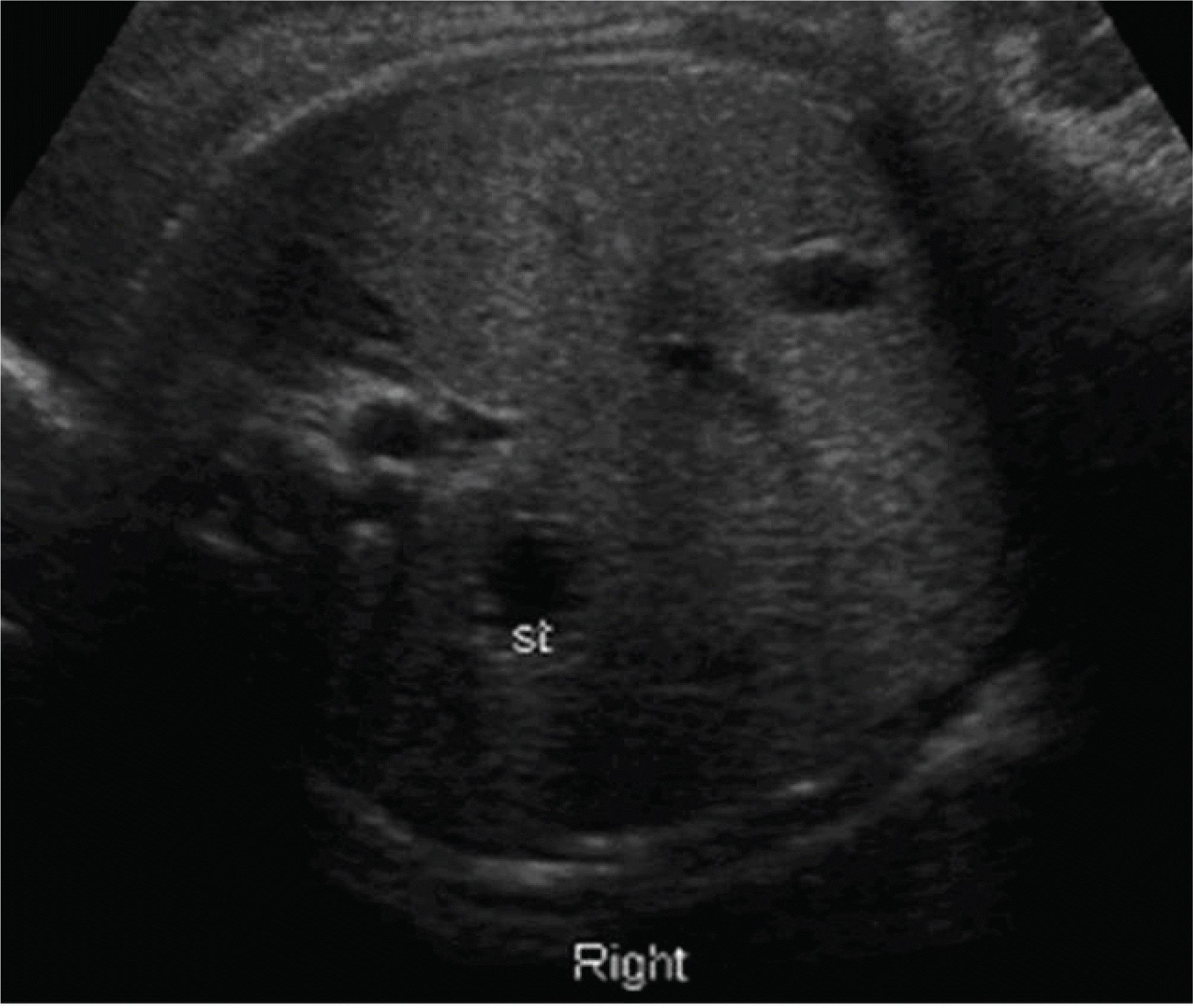
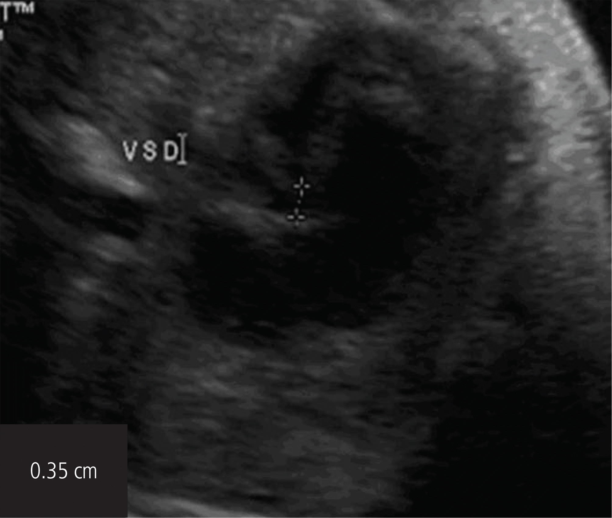
Fig. 2.
Prenatal sonography showed abnormal findings. Right ventricle larger than left ventricle in a ratio of 1.62 cm:0.63 cm with ventricular septal defect. VSD, ventricular septal defect.
 XML Download