Abstract
Extramammary Paget's disease is a rare, slow-growing, non-invasive intraepithelial adenocarcinoma that occurs mainly in the elderly outside of the mammary gland. Paget's disease of the vulva accounts for less than 1% of all vulvar malignancies. The standard treatment is wide local excision of the gross lesion. To prevent a relapse, wide excision with adequate margins is necessary not to leave pathologic lesions around margins of excision. However, since the excised area is too wide to cover all the exposed surface with a simple closure only, skin grafts and flaps can be very useful. We report successful treatment of three cases of extramammary Paget's disease of the vulva using frozen section analyses of the margins during operation in order to attain Paget's cells free margins and skin grafts and flaps to cover an extensive defects.
REFERENCES
1. Black D, Tornos C, Soslow RA, Awtrey CS, Barakat RR, Chi DS. The outcomes of patients with positive margins after excision for intraepithelial Paget's disease of the vulva. Gynecol Oncol. 2007; 104:547–50.

2. Tebes S, Cardosi R, Hoffman M. Paget's disease of the vulva. Am J Obstet Gynecol. 2002; 187:281–3.

3. Wilkinson EJ, Brown HM. Vulvar Paget disease of urothelial origin: a report of three cases and a proposed classification of vulvar Paget disease. Hum Pathol. 2002; 33:549–54.

4. Fanning J, Lambert HC, Hale TM, Morris PC, Schuerch C. Paget's disease of the vulva: prevalence of associated vulvar adenocarcinoma, invasive Paget's disease, and recurrence after surgical excision. Am J Obstet Gynecol. 1999; 180:24–7.

5. Yoon MK, Jeon YE, Park YH, Kim SJ, Kang JB, Jang BR, et al. A case of vulvar adenocarcinoma associated with extramammary Paget's disease. Korean J Obstet Gynecol. 2006; 49:939–44.
6. Feuer GA, Shevchuk M, Calanog A. Vulvar Paget's disease: the need to exclude an invasive lesion. Gynecol Oncol. 1990; 38:81–9.

7. Kanitakis J. Mammary and extramammary Paget's disease. J Eur Acad Dermatol Venereol. 2007; 21:581–90.

9. Curtin JP, Rubin SC, Jones WB, Hoskins WJ, Lewis JL Jr. Paget's disease of the vulva. Gynecol Oncol. 1990; 39:374–7.

10. Fishman DA, Chambers SK, Schwartz PE, Kohorn EI, Chambers JT. Extramammary Paget's disease of the vulva. Gynecol Oncol. 1995; 56:266–70.

11. Kodama S, Kaneko T, Saito M, Yoshiya N, Honma S, Tanaka K. A clinicopathologic study of 30 patients with Paget's disease of the vulva. Gynecol Oncol. 1995; 56:63–70.

12. Parker LP, Parker JR, Bodurka-Bevers D, Deavers M, Bevers MW, Shen-Gunther J, et al. Paget's disease of the vulva: pathology, pattern of involvement, and prognosis. Gynecol Oncol. 2000; 77:183–9.

13. Roh HJ, Kim DY, Kim JH, Kim YM, Kim YT, Nam JH. Paget's disease of the vulva: evaluation of recurrence relative to symptom duration, volumetric excision of lesion, and surgical margin status. Acta Obstet Gynecol Scand. 2010; 89:962–5.

14. Geisler JP, Stowell MJ, Melton ME, Maloney CD, Geisler HE. Extramammary Paget's disease of the vulva recurring in a skin graft. Gynecol Oncol. 1995; 56:446–7.

15. Tjalma WA, Cooremans ID, Jeuris W, Van Marck EA, Monaghan JM. Recurrent Paget's disease of the vulva in a myocutaneous flap: case report and review of the literature. Eur J Gynaecol Oncol. 2001; 22:13–5.
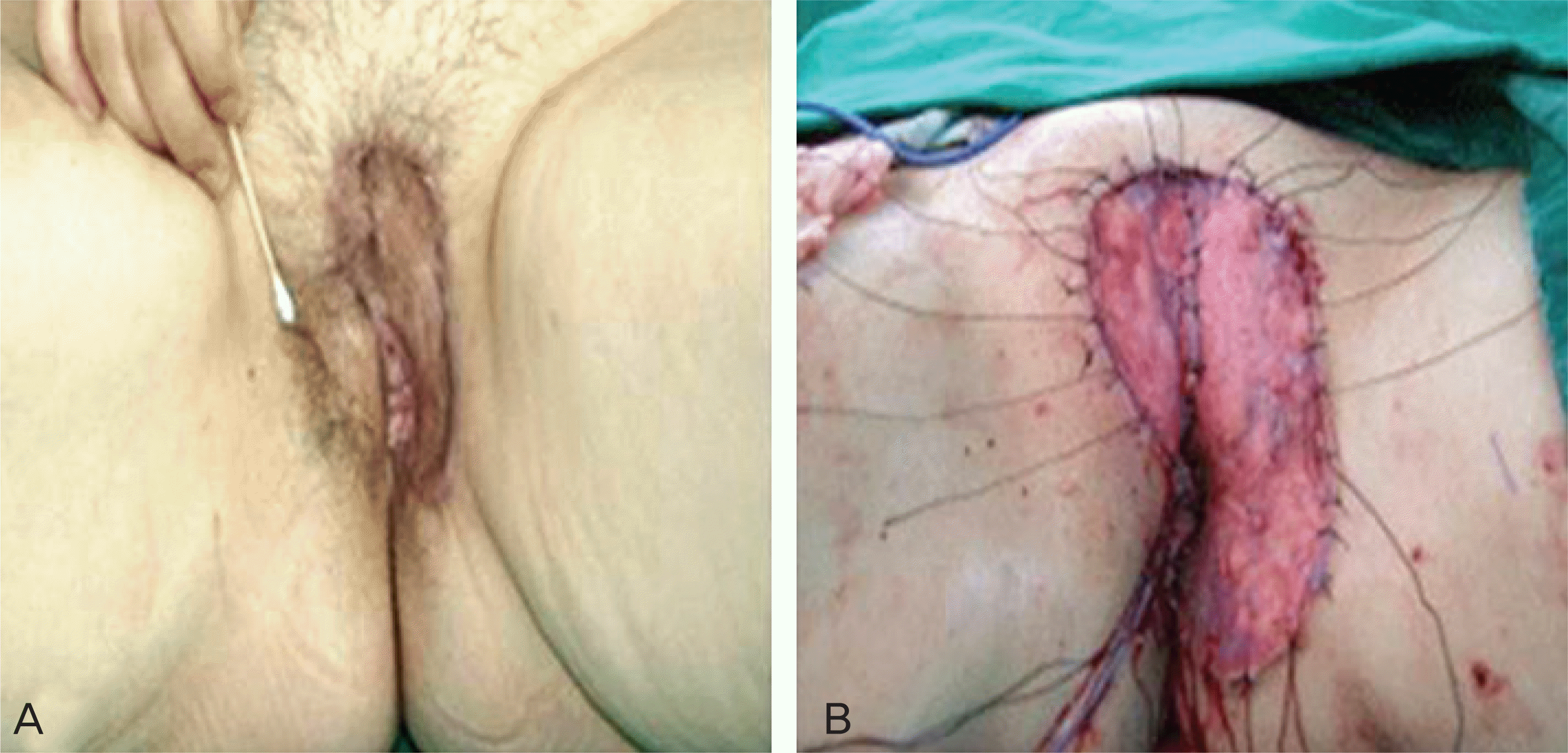
Fig. 1.
Preoperative finding: eczematoid lesion on left labium major (A). Postoperative finding: wide local excision and coverage by split-thickness skin graft (B).

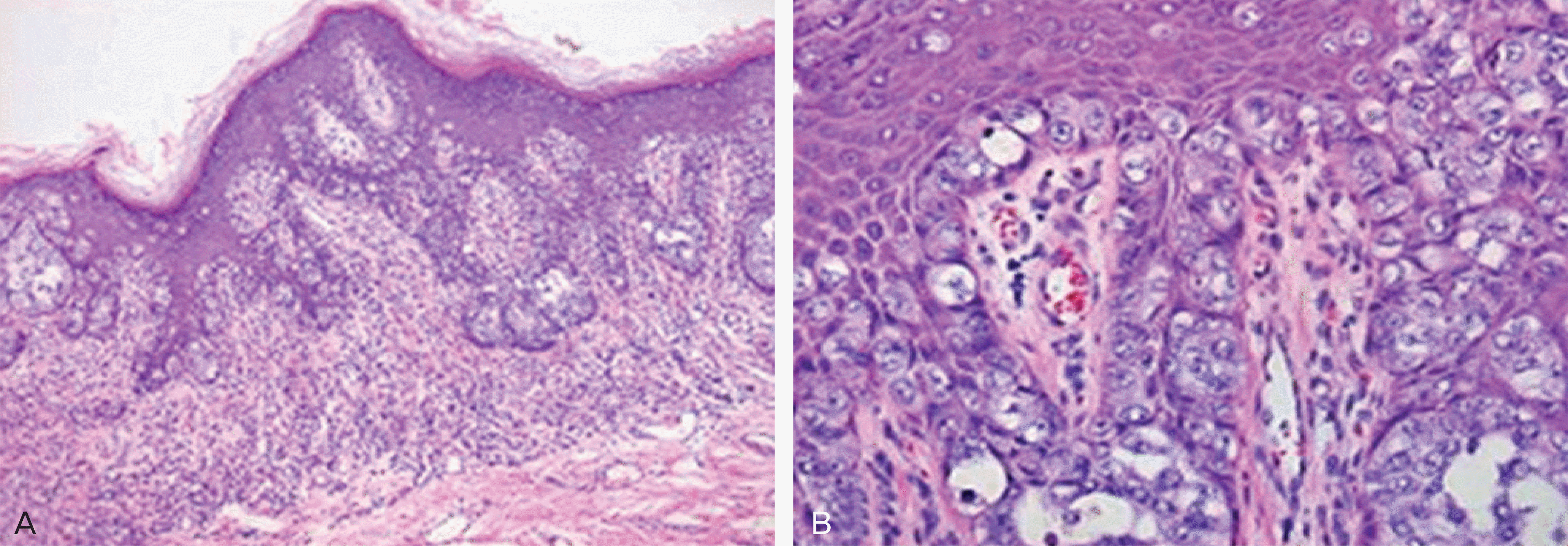
Fig. 2.
Microscopic features of vulvar Paget's disease. Large pale tumor cells dispersed singly or in clusters of solid nests and glandular spaces within the epidermis (H&E, ×100) (A). Tumor cells have abundant pale cytoplasm with large nucleus and prominent nucleolus at high-power view (H&E, ×400) (B).


 PDF
PDF ePub
ePub Citation
 Print
Print


 XML Download