

 PDF
PDF ePub
ePub Citation
 Print
Print


Endometriosis, one of the most common gynecologic disorders, is broadly defined as the presence of endometrial glandular and stromal cells outside the uterine cavity, associated with symptoms of dysmenorrhea, dyspaureunia, chronic pelvic pain and subfertility. But, the etiology and pathogenesis of endometriosis remain obscure. Theories that account for this susceptibility include genetic predisposition [1], large amount of retrograde menstruation [2], an altered peritoneal environment [3], or an immunological susceptibility [4]. The most widely accepted theory is that the disease is caused by retrograde menstruation and subsequent implantation of endometrial glands on the surface of the abdominal cavity [2,5]. However, retrograde menstruation is frequently observed in women unaffected by endometriosis and such menstrual debris does not result in endometriosis in all women. Thus, additional factors may be present in the uterine endometrium of women who have developed the disease. The women who develop endometriosis are due to abnormalities inherent to their ectopic or eutopic endometrium.
The refluxed menstrual debris in women with endometriosis may be more prone to implant, invade and grow in peritoneum or ovary through the action of extracellular proteolysis and angiogenesis. Angiogenesis is facilitated by proteolysis, since endothelial cells require proteolytic activity to be able to degrade their basal membrane, to migrate and to invade the underlying extracellular matrix [6-8]. Key regulators of proteolysis belong to the family of matrix metalloproteinases (MMPs). They represent a large family of proteolytic enzymes regulated by tumor-stromal interaction that play key roles in cancer progression, promoting proliferation, angiogenesis and tumor metastasis [9]. In particular, the membrane-type matrix metalloproteinases (MT-MMPs) are a new subfamily of membrane-anchored MMPs, which as of today includes six members: MT1-, MT2-, MT3-, MT4-. MT5-, and MT6-MMP. Among them, MT1-, MT2-, MT3-, and MT5-MMPs are trans-membrane proteins. Their membrane-associated localization makes them particularly suited to functioning in pericellular proteolysis [10,11]. Previous reports showed that MT-MMPs play an important role in angiogenesis [10,12,13], especially MT1-MMP has received considerable attention as being involved in tumor angiogenesis [14,15]. Several MT-MMPs have been demonstrated in whole endometrial extracts at mRNA level [16,17], and MT1- and MT2-MMP antigens have been demonstrated in various endometrial cell types [18,19].
MT-MMPs act at the cell surface where they can locally facilitate degradation of extracellular matrix, cell migration, invasion and angiogenesis. The abundance of all MT-MMP in cycling endometrium suggests that endometrial MT-MMPs play a role in remodeling of cycling endometrium in preparation for implantation [20]. MT-MMPs are inhibited by tissue inhibitor of matrix metalloproteinase-2 (TIMP-2) [16]. The endometriosis-associated increase in proteolysis and imbalance between the secretion of MMP-9 and that of its natural inhibitor, TIMP-1, revealed in the culture medium of endometrial tissue [21]. The possible changes in MT-MMPs activity in the eutopic endometrial tissue of patients with endometriosis suggest an enhanced proteolysis which could play a role in enabling this tissue to implant in ectopic locations.
Therefore, the aim of the present study was to investigate whether the endometrial tissue from women with endometriosis would express a higher MT2-MMP and MT3-MMP mRNA expression consistent with higher angiogenic activity and increased growth.
Materials and Methods
1. Tissue collection
Endometrial samples were obtained from 79 premenopausal women aged 29-45 years, undergoing laparoscopic surgery or hysterectomy for non-malignant lesions. Patients with pelvic inflammatory disease, adenomyosis and dysfunctional uterine bleeding were excluded. Patients have not taken the nonsteroidal anti-inflammatory drugs, GnRH agonists and steroids for the past 6 weeks. Sufficient eutopic endometrial tissues were available from 36 patients with endometriosis stages III and IV endometriosis diagnosed by both pathology and laparoscopic findings according to the revised American Fertility Society classification of endometriosis [22]. Endometrial tissue from 52 control patients without endometriosis confirmed by laparoscopic surgery was also collected. The study protocol was approved by the Institutional Review Board on the Use of Human Subjects in Research at Ewha Womans University and informed consent was obtained.
Endometrial samples were taken using a pipette in the operating room before the laparoscopic procedure; in patients undergoing hysterectomy, the uterine cavity was opened and endometrium obtained immediately after the specimen was removed. Tissue samples were classified by histological dating according to the method of Noyes et al. [23] into two groups: proliferative phase (n=49) and secretory phase (n=30). The remaining tissue was washed in PBS solution in order to remove contaminating blood and RNA was immediately extracted.
2. RNA extraction
The extraction of RNA from the tissue sample was carried out with the RNA-STAT-60 reagent (Tel-Test "B" Inc., Friendswood, TX, USA). Briefly, tissue samples were washed three times in PBS (Gibco BRL, Grand Island, NY, USA) to remove blood contamination. One hundred milligrams of tissue were homogenized in 1 mL of RNA-STAT-60 reagent. Total RNA was separated from DNA and proteins by adding chloroform and was precipitated using isopropanol. The precipitate was washed two times in 75% ethanol, air-dried, and re-diluted in diethylpycocarbonate (DEPC)-treated dH2O. The amount and purity of extracted RNA was quantitated by spectrophotometry in a GenQuant RNA/DNA calculator (Pharmacia Biotech Ltd., Cambridge, UK) and 10-100 µg of total RNA was routinely obtained.
3. Reverse transcription (RT) PCR
Specific sequences of oligonucleotide primers for MT2-, and MT3-MMP were obtained from Gene Bank Database of the National Center for Biotechnology Information of the National Institutes of Health (NIH, Internet address: http://www.2.ncbi.nlm.nih.gov/cgibin/genbank). One corresponding set of primers for MT2-MMP and MT3-MMP was found with the help from the program OLIGO 5.0 Primer Analysis Software (National Bioscience, Plymouth, MN, USA) and synthesized by Biomed, Seoul, Korea. The primer sequences, locations on the mRNA, and sizes of the amplified fragments are listed in Table 1.
For RT-PCR, the Gen Amp RNA PCR kit (Perkin-Elmer, Foster City, CA, USA) was used. Nineteen microliters of RT-mastermix for each sample were prepared containing 5 mmol/L MgCl2, 1X PCR buffer II, 1 mmol/L of each deoxy-NTP, 2.5 µL/L oligo (deoxythimidine)16, 20 IU ribonuclease inhibitor (all from Perkin-Elmer), 100 IU Moloney murine leukemia virus reverse transcriptase (Gibco BRL), and 1 µg total RNA diluted in 1 µL DEPC-treated H2O and placed into 0.2 mL thin wall PCR tube (Applied Scientific, South San Francisco, CA, USA). RT was carried out in the DNA Thermal Cycler 9600 (Perkin-Elmer) using a program with the following parameters: 42℃, 15 min; 99℃, 5 min; then quenched at 4℃. After the reaction was completed, samples were stored at -20℃ until the PCR. As a negative control, 1 µL DEPC-treated H2O without RNA sample was subjected to the same RT reaction.
4. Construction of the competitive and target cDNA fragment for MT2-, and MT3-MMP
A 428 base pair (bp), and 532 bp fragment of native MT2-MMP, and MT3-MMP cDNA (the target) were obtained by PCR amplification of reverse-transcribed total RNA from endometrial biopsies with the regular 3' and 5' primers (Table 1). The PCR product was visualized by agarose gel electrophoresis stained with ethidium bromide (EtBr), cDNA was extracted from the gel, purified with an agarose gel extraction kit (Amersham Pharmacia Biotech Ltd., Piscataway, NJ, USA) and quantitated by spectrophotometry (Pharmacia Biotech Ltd., Cambridge, UK).
To construct a competitive cDNA fragment, a floating primer with a sequence complementary to cDNA between the 3' and 5' primer binding sites was designed by attaching the complementary sequence of the binding site of the original 3'-MT2-MMP, and MT3-MMP. After PCR with the regular 5'-primer and the 3'-floating primer, the PCR product was visualized by agarose gel electrophoresis stained with EtBr. cDNA extraction, purification, and concentration determination were performed as described above. These steps resulted in cDNA fragments of 201 bp and 415 bp, each with 3'-end and 5'-end primer binding sites on their ends which were products of 227 bp and 117 bp deletion from the target cDNA, respectively.
5. Standard curve and competitive PCR for MT2-, and MT3-MMP
The standard curve for MT2-MMP, and MT3-MMP was constructed by co-amplification of the constant amount of competitive cDNA (0.1 fmol for MT2-MMP, and 1 fmol for MT3-MMP ) with declining amounts of target cDNA (from 62.5 to 0.01525 fmol for MT2-MMP and from 500 to 0.122 fmol for MT3-MMP ) obtained by serial dilution. A total of 100 µL of PCR mixture containing 1.9 mmol MgCl2 solution, 10X PCR buffer II, 0.2 mmol/L of deoxy-NT, and 2.5 U Taq-polymerase (all from Perkin-Elmer) with corresponding paired primers at a concentration of 0.2 µmol/L of each primer, was placed in the Perkin-Elmer DNA Thermal Cycler 9600. PCR cycles were composed of 1 cycle of 95℃ for 5 min to denature all proteins, 35 cycles of 45 sec at 94℃, 45 sec at 57℃, and 45 sec at 72℃ for MT2-MMP, and 30 cycles of 45 sec at 94℃, 45 sec at 62℃, and 45 sec at 72℃ for MT3-MMP. The reaction was terminated at 72℃ for 7 min and was quenched at 4℃. One percent agarose gel electrophoresis was carried out in a H5 electrophoresis chamber. Gels were stained with EtBr. Aliquots (25 µL) of each PCR product and dye buffer were analyzed in parallel with a 100 bp DNA ladder as a standard.
After completion of electrophoresis, the gel blot was analyzed and photocopies of the blot were printed by UV densitometry (Gel-Doc and Chemidoc system, Bio-Rad Laboratories, Hercules, CA, USA). The logarithmically transformed ratios of target cDNA to competitive cDNA were plotted against the log amount of initially added target cDNA in each PCR to obtain linear and reproducible standard curves. Values obtained from the regression line of the standard curve (y=b+mx) allowed us to calculate the amount of cDNA transcripts in an unknown sample: 0.1 fmol of MT2-MMP, and 1 fmol of MT3-MMP competitive cDNA were added to each unknown sample before PCR. The ratio of the densities of sample target cDNA band (428 bp, and 532 bp) to competitive cDNA (201 bp and 415 bp) were logarithmically transformed and compared the values obtained from standard curve. Quantitative competitive PCR was carried out on at least two aliquots from the RT cDNA of each patient, and the results did not differ more than ±5% and we used the average concentration for data analysis.
Results
1. RT-PCR of endometrial tissue throughout the menstrual cycle
RT-PCR was employed to increase the sensitivity of detection, and the 838 bp sequence of β-actin, 428 bp sequence of MT2-MMP, and 532 bp sequence of MT3-MMP mRNA were expressed by all eutopic endometrial samples from women with and without endometriosis in both the proliferative and secretory phase of the menstrual cycle. β-actin mRNA expression was also measured in all the samples studied, thus confirming the integrity of RNA and the RT-PCR process (data not shown).
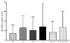
2. Quantitative MT2-MMP mRNA expression in eutopic endometrial tissue from women with or without endometriosis
Throughout the menstrual cycle, eutopic endometrium from patients with endometriosis did not show differences of MT2-MMP mRNA expression compared to eutopic endometrium from control (Fig. 1).
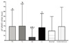
3. Quantitative MT3-MMP mRNA expression in eutopic endometrial tissue from women with or without endometriosis
Quantitative expression of MT3-MMP mRNA in eutopic endometrium of patients with endometriosis was higher in secretory phase compared to that of control group. During the proliferative phase, there are no differences between eutopic endometrium with and without endometriosis (P > 0.05) (Fig. 2).
Discussion
MT-MMPs are of interest in pathogenesis of endometriosis because of their specific features involving matrix degradation and activation of other MMPs. It is understood that the tissue destruction and invasion in endometriosis are mediated by the concerted action of various proteinases, among which the MMPs appear to play a major role. Recently, novel members of the MMP family, the MT-MMPs, have been described [24-28]. It is of particular importance that at least three of these MT-MMPs, namely, MT1-, MT2-, and MT3-MMP, have been shown to be capable of not only degrading extracellular matrix components but also activating other MMPs, such as MMP-2 and MMP-13 [24,25,29-31]. MT-MMPs (with the exclusion of MT4-MMP ) activate MMP-2 (gelatinase A), an enzyme that has a key role in local invasion and dissemination of a large variety of tumors [24,32-34], through a 2-step cleavage reaction, following the formation of a membrane complex with MMP-2 and tissue inhibitor of metalloproteinase-2 (TIMP-2) [32]. The MT-MMPs are inhibited by TIMPs family which includes four members (TIMP-1, TIMP-2, TIMP-3, and TIMP-4). For example, MT1-, MT2-, MT3-, and MT5-MMP are efficiently inhibited by TIMP-2 and TIMP-3. The efficiency in mediating this cleavage reaction seems to be highest for MT1-MMP, followed by MT3-MMP, and is lower for MT2-MMP [34]. MT1-MMP also can activate procollagenase-3 (MMP-13), while recombinant forms of all 3 enzymes can cleave a large number of ECM proteins [34].
In this study, the eutopic endometrium from women with endometriosis expressed higher levels of MT3-MMP than that from normal women. But MT2-MMP expression from eutopic endometrium showed no significant differences between patients with endometriosis and controls.
Several studies have suggested a role for MT-MMPs in the degradation of the extracellular matrix in malignancies and rheumatoid arthritis [35-39]. MT1-MMP may play a key role in human breast carcinoma invasion and metastasis [35]. In the other study, it was found that the expression of MT-MMP in cervical cancer cells both in vitro and in vivo was higher in invasive cervical carcinoma and lymph node metastases compared to its expression in non-invasive CIN III lesions [36]. Another study found a higher level of MT-MMP expression that MT1-MMP and MT2-MMP play an important role in the development of human urothelial carcinomas [37]. A role for MT1-MMP is not only in the matrix degradation by fibroblasts, but also in osteoclast-mediated bone resorption in RA [38]. MT1-MMP and MT2-MMP were able to directly confer invasion-incompetent cells with the ability to penetrate type I collagen matrices. MT-MMP-expressing cells can penetrate and remodel type I collagen-rich tissues by using membrane-anchored metalloproteinases as pericellular collagenases [10].
Previous data presented suggested a positive correlation between expression of MMP-2, MT1-MMP, and MT2-MMP mRNA and, possibly, a role in ovarian carcinoma and endometriosis pathogenesis, mainly through tumor cell production of these enzymes [35,39]. So, we compared the patients with endometriosis and controls in expression of MT-2 and MT3-MMP.
All MT-MMPs are expressed in endometrium in a cycle-dependent pattern with decreased levels during the early secretory phase. MT-MMPs may play a role in endometrial remodeling in preparation for implantation and were reduced during the receptive window [20]. And in our study also eutopic endometrium from control have tendency lower expression levels in secretory phase compare than proliferative phase.
In this study, MT-3 MMP is related with proteolysis and angiogenesis of endometriosis. It suggests that increased ability to proteolysis and angiogenesis of endometrium is essential for surviving outside the uterus in endometriosis. In conclusion, the MT-MMPs system may play an important role in the pathogenesis of endometriosis.

 XML Download