

 PDF
PDF ePub
ePub Citation
 Print
Print


The physiological changes occurring during pregnancy and the process of childbirth have a detrimental effect on the structure and function of the muscles, nerves, and fascial tissues (connective tissues) that make up the pelvic floor complex. As the fetus grows, the weight of the fetus and gravid uterus produce various anatomic changes.
The levator ani muscle is believed to play an important role in supporting the pelvic organs and maintaining normal pelvic floor function [1-4]. Anatomically, the levator ani muscle is composed of two portions, the lateral supportive iliococcygeus and the central sphincteric puborectalis and pubococcygeus (or pubovisceral muscles) [1,2,4]. The levator ani muscle also undergoes hypertrophy during the course of pregnancy [5].
Imaging of the pelvic floor muscles, to further determine their role in support of the pelvic organs, continence, and childbirth, has been a focus of research in recent years. Changes in the morphology of the pelvic floor following vaginal delivery, incontinence, and prolapse have been demonstrated using magnetic resonance imaging (MRI) and, more recently, three dimensional (3D) ultrasound [1,6-8]. To date, MRI has been the imaging method of choice due to its superior spatial resolution capabilities and, as shown most recently [9], its ability to identify different muscle groups of the pelvic floor. However, recent technological advances in 3D ultrasound have allowed access to the axial plane and direct imaging of the entire levator hiatus, previously the domain of MRI. Several studies have recently defined characteristics of the pelvic floor following delivery, especially, the effects of delivery mode on the pelvic floor [10,11]. However, there is still a lack of studies describing the effects of pregnancy itself on the pelvic floor.
The aim of this study was to evaluate morphological characteristic of the pelvic floor in pregnant women using 2D- and 3D-transperienal ultrasound and compare with non-pregnant women.
Materials and Methods
This was a case-control study of pregnant and non-pregnant women visiting the Guro Hospital, Korea University Medical Center from November, 2009 to March, 2010. The study population consisted of 40 nulliparous pregnant women at term and 28 age matched nulliparous non-pregnant women.
1. Study participants
The case group included pregnant women who had singleton vertex fetus. The control group included non-pregnant, age matched women who visited the hospital for routine gynecological checkups and had no specific findings on ultrasound. Women who had a previous gynecologic surgery, especially myomal and adnexal surgery, such as for endometriosis and adhesiolysis or history of fecal or urinary incontinence, were excluded as these conditions may influence pelvic floor function. Women who had a complicated pregnancy or had difficulties due to poor cooperation were also excluded.
All subjects provided written informed consent prior to participation in the study, which was approved by the ethical committee at our institution. The patients underwent a semi-structured interview about past history and continence status.
2. Measurements on ultrasound
The images presented here were obtained by transperineal ultrasound performed with the same equipment and examiner. The evaluation was performed with low pressure, just sufficient to obtain an image with good resolution.
According to the German Association of Urogynecology, there are three methods for the measurement of bladder neck position. The first is the measurement of two distances and the second is the measurement of one distance and one angle. The third is the measurement of the height of the bladder neck with a horizontal line is drawn at the lower border of the symphysis. The third method provides reliable results only if the position of the ultrasound probe is stable. The current study used the one distance and one angle method for measurement of the bladder neck position [12].
1) 2-dimensional ultrasound measurements
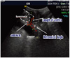
Measurements of the distance between the bladder neck and the lower border of the symphysis (dBNS) and the angle between this distance line and the central line of the symphysis (retro-vesical β-degree) were obtained by transperineal 2D ultrasound (Fig. 1) [11,12]. The length of the urethra was determined as the distance between bladder neck and horizontal line at the lower border of symphysis by transperineal 2D ultrasound [13]. Bladder volume and bladder wall thickness have minimal effects on the distance and angle measurements [14-16]. However, according to Dietz, mobility of the bladder neck is increased when the bladder is empty [16,17]. The bladder volume and bladder wall thickness was measured as it related to detrusor overactivity and urinary incontinence [16,18].
Bladder volume was measured with three parameters, including height (H), depth (D), and width (W). These measures were obtained from perpendicular planes (sagittal and transverse) by transperineal 2D ultrasound. In sagittal scanning, height and depth correspond to the greatest superior-inferior measurement and the greatest anterior-posterior measurement, respectively. Thus, the bladder volume (mL)=H × D × W × 0.7. The value of 0.7 is a correction factor for the nonspherical shape of a full bladder. This examination was performed as the bladder neck position is influenced by bladder volume. The bladder wall thickness was measured as a hypoechoic layer sandwiched between two hyperechoic layers, the urothelium and perivesical tissue [19].
2) 3-dimensional ultrasound measurements
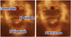
We checked the maximum diameter of the levator hiatus (anteroposterior [AP] diameters and transverse diameters) in mid-sagittal imaging and measured the maximum diameters of the pubovisceral muscle thickness (left and right of the rectum) at the level of maximal muscle thickness by 3D transperineal ultrasound (Fig. 2) [10,13]. The pubovisceral angle is the line between the vertical line that transverses the urethra and rectum and the tangent line that connects the point where the vertical line crosses the inner edge of the puborectal loop with the most distant inner point of the pubovisceral muscle [20].
To obtain reliable results with this method, we carried out measurements in the semi-supine position, after voiding, while at rest and during the Valsalva maneuver. The one specialist sonographer was assessed total ultrasounds were carried out by a radiologic technician. A Medison Accuvix XQ Ultrasound system (Medison Healthcare, Seoul, Korea) with a 4-7 MHz curved array ultrasound transducer was used for all measurements.
Results
Satisfactory biometric measurements were obtained for 40 nulliparous pregnant women at term and 28 nulliparous non-pregnant women.
Table 1 shows basic characteristics of study participants according to pregnancy status. There were no significant differences in age and height between groups. The mean body mass index (BMI) was higher in the pregnant group compared to the non-pregnant group (29.10 ± 3.27 kg/m2 vs. 22.04 ± 3.50 kg/m2; P ≤ 0.001).
There was a no difference between pre-pregnant BMI and non-pregnant BMI (22.22 ± 3.00 kg/m2 vs. 22.04 ± 3.50 kg/m2; P ≤ 0.537).
The characteristics of the pelvic floor assessed by 2D ultrasonography between pregnant and non-pregnant women are summarized in Table 2. The length of urethra was longer in pregnant women at rest and during the valsalva maneuver compared with non-pregnant women (3.54 ± 0.62 cm vs. 2.70 ± 0.73 cm; P ≤ 0.007) and (3.32 ± 0.57 cm vs. 2.55 ± 0.47 cm; P=0.005), respectively. The distance between the dBNS was shorter in pregnant women at rest and during the valsalva maneuver compared with non-pregnant women (0.68 ± 0.24 cm vs. 1.07 ± 0.30 cm; P=0.002 and 0.49 ± 0.11 cm vs. 0.99 ± 0.38 cm; P ≤ 0.001), respectively. The retro-vesical β-degree, bladder thickness, and bladder volume were not significantly different between pregnant and non-pregnant women, both at rest and during the valsalva maneuver (P ≥ 0.05).
Table 3 shows the characteristics of the levator hiatus obtained by 3D transperineal ultrasonography according to pregnancy status. The anteroposterior diameter of the levator hiatus was not significantly different between pregnant women and non-pregnant women (P=0.423). The mean thickness of the pubovisceral muscle was greater in pregnant than non-pregnant women (1.03 ± 0.15 cm vs. 0.77 ± 0.16 cm; P=0.001) (Fig. 3). The transverse diameter of the levator hiatus and the pubovisceral angle of non-pregnant women were significantly greater than pregnant women. (3.97 ± 0.67 cm vs. 3.45 ± 0.49 cm; P=0.033 and 60.67° ± 9.68° vs. 51.75° ± 8.52°; P=0.026) (Figs. 4, 5).
Discussion
To our best knowledge, this is the first study to evaluate morphological characteristic of the pelvic floor in pregnant women using 2D- and 3D-transperienal ultrasound. Pregnant women had a significantly greater thickness of the pubovisceral muscle and decreased transverse diameter and pubovisceral angle. These changes reflect the fact that pregnant women have a smaller hiatal area than non-pregnant women. Morphological changes in the levator hiatus may have clinical significance in the subsequent development of urinary incontinence and pelvic organ prolapsed [16,21]. The levator ani muscle is thought to play a significant role in the pathogenesis of these highly prevalent conditions and it is estimated that parous women have an increased lifetime risk (by age 80) of undergoing surgical treatment for one of these conditions [22]. Pregnancy and childbirth are frequently cited as major etiological factors, and various obstetric parameters (e.g., length of second-stage labor, birth weight, and mode of delivery) have been demonstrated to be additional risk factors [23,24]. A potential protective effect of Cesarean section could not be verified in long-term studies, suggesting that pregnancy itself (especially the first) causes pathological changes to the pelvic floor, regardless of the mode of delivery [24-26]. It has been suggested that the strain of the gravid uterus and hormonal changes during pregnancy lead to connective tissue remodeling and disruption of normal pelvic floor function; additional disruption may result during vaginal delivery from traumatic damage, primarily by vacuum or forceps extraction [27].
It is interesting to note that the hiatal area at term was decreased. Although the reason for this change is unclear, a possible explanation is that during pregnancy, the volume and weight of the uterus increase and the pressure of the uterus and fetal presentation on the pelvic floor also increases [28]. Therefore, the decreased hiatal area may reflect a compensatory mechanism counteracting these changes. Pregnancy itself may cause morphological changes of the pelvic floor to support the birth canal by closing the lower end of the pelvic cavity as diaphragm. However, these changes may have some detrimental effects in pregnancy. During vaginal delivery, muscles of the levator hiatus are required to deform and stretch markedly. Using computer modeling, based on MRI, it has been found that some parts of the pubovisceral muscle stretch to 3.3 times their resting length during crowning of the head [29]. It is unlikely that the pelvic floor presents an absolute barrier to descent and delivery of the presenting part in any but a very small number of women. However, in the context of modern obstetric practice, a less compliant pelvic floor may provoke intervention by prolonging the second stage, slowing descent of fetal presentation, and predisposing to fetal distress. Interestingly, in a recent study of pelvic floor training in pregnancy, pelvic floor exercises were found to decrease the incidence of a prolonged second stage [30].
Therefore, further studies are needed to understand how the changes to the pelvic floor during pregnancy may influence the progress of labor. We found the distance between the bladder neck and the inferior border of the symphysis, both at rest and during the Valsalva maneuver, were significantly decreased in pregnant women compared with non-pregnant women. There was an increase in the length of the urethra, both during at rest and the Valsalva maneuver, in pregnant women.
Modification of the anatomic relationship between the bladder and enlarged uterus results in shortening of the distance between the dBNS and increasing the length of the urethra.
Recent technological advances in ultrasound imaging have resulted in three-dimensional pelvic floor imaging that is able to demonstrate the levator muscle in the axial plane in a manner comparable to MRI [31]. The facility of real-time data capture also permits examination of functional anatomy observed during the Valsalva maneuver and pelvic floor muscle contraction. These factors, as well as its relatively low cost and associated high patient compliance, mark ultrasound as an ideal method for further study of the levator in pregnancy [32].
In conclusion, pregnant women had significantly thicker levator ani muscles but smaller hiatal areas, as measured by the levator hiatus angle and transverse diameter, than did non-pregnant women. Pregnancy itself may cause morphological changes of pelvic floor to support the birth canal by closing the lower end of the pelvic cavity as a diaphragm.


 XML Download