

 PDF
PDF ePub
ePub Citation
 Print
Print


Abstract
Objective
To investigate the association between the cervical angiogenin and interleukin-6 levels and its outcome during early pregnancy.
Methods
One hundred and seventy-five pregnant women were included consecutively at the time of their first prenatal visit after less than 10 weeks of gestation. The gestational age was calculated according to their menstrual history and/or ultrasonography. We measured the cervical angiogenin and interleukin-6 levels using ELISA kits. We also measured the maternal serum progesterone concentrations using radioimmunoassay. Spontaneous abortion was defined as when a pregnancy ended spontaneously within 20 weeks of gestation. If an index pregnancy lasted longer than 20 weeks of gestation, it was regarded as showing a normal pregnancy outcome.
Results
Fifty-four pregnancies were excluded because of an incomplete study (n=36) or follow-up loss (n=18). Of the remaining 121 pregnancies, spontaneous abortion occurred in 10 pregnancies (8.3%) in which there were no significant gestational age-dependent changes in the maternal serum progesterone, cervical angiogenin, and interleukin-6 levels. The maternal serum progesterone, cervical angiogenin, and interleukin-6 levels were not significantly different between the spontaneous abortion group and the normal control group. However, the cervical angiogenin level was significantly decreased in the groups with discordant gestational age and spontaneous abortion compared to the group with concordant gestational age (median, 798.0 pg/mL vs. 723.4 pg/mL vs. 1039.8 pg/mL; inter-quartile range, 222.8-1054.7 pg/mL vs. 257.1-1213.3 pg/mL vs. 480.4-1534.4 pg/mL) (p<0.041).
Figures and Tables
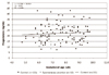 | Fig. 1Scattergram of maternal serum progesterone levels in normal (♦; n=111) and spontaneous abortion group (▪; n=10). Note the values of spontaneous abortion group are scattered evenly up and below the gestational age-related trend line for the values of normal group.
|
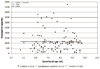 | Fig. 2Scattergram of cervical angiogenin levels in normal (♦; n=111) and spontaneous abortion group (▪; n=10). Note the most values of spontaneous abortion group are scattered below the gestational age-related trend line for the values of normal group.
|
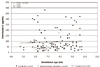 | Fig. 3Scattergram of cervical interleukin-6 levels in normal (♦; n=111) and spontaneous abortion group (▪; n=10). Note the most values of spontaneous abortion group are scattered below the gestational age-related trend line for the values of normal group.
|
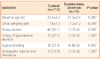
Table 2
Maternal serum progesterone and cervical angiogenin and interleukin-6 levelsa in normal and spontaneous abortion group

![]()
Table 3
Clinical characteristics of control group stratified by discordancya in gestational age calculated by menstrual period versus ultrasonography

![]()
Table 4
Maternal serum progesterone and cervical angiogenin and interleukin-6 levelsa in the control group stratified by discordancyb in gestational age calculated by menstrual period versus ultrasonography

![]()

References
1. Scott JR. Scott JR, DiSaia PJ, Hammond CB, Spellacy WN, editors. Early pregnancy loss. Danforth's obstetrics and gynecology. 1999. 8th ed. Philadelphia: Lippincott Williams & Wilkins;143–153.
2. Batzofin JH, Fielding WL, Friedman EA. Effect of vaginal bleeding in early pregnancy on outcome. Obstet Gynecol. 1984. 63:515–518.
3. Funderburk SJ, Guthrie D, Meldrum D. Outcome of pregnancies complicated by early vaginal bleeding. Br J Obstet Gynaecol. 1980. 87:100–105.
4. Fossum GT, Davajan V, Kletzky OA. Early detection of pregnancy with transvaginal ultrasound. Fertil Steril. 1988. 49:788–791.
5. al-Sebai MA, Kingsland CR, Diver M, Hipkin L, McFadyen IR. The role of a single progesterone measurement in the diagnosis of early pregnancy failure and the prognosis of fetal viability. Br J Obstet Gynaecol. 1995. 102:364–369.
6. Kim JI, Kim KJ, Hwang WJ, Han JS, Won HS, Lee PR, et al. The value of a single progesterone measurement in the predictor of early pregnancy failure and the prognosis of fetal viability. Korean J Perinatol. 1996. 7:304–308.
7. Strydom DJ, Fett JW, Lobb RR, Alderman EM, Bethune JL, Riordan JF, et al. Amino acid sequence of human tumor derived angiogenin. Biochemistry. 1985. 24:5486–5494.
8. Fett JW, Strydom DJ, Lobb RR, Alderman EM, Bethune JL, Riordan JF, et al. Isolation and characterization of angiogenin, an angiogenic protein from human carcinoma cells. Biochemistry. 1985. 24:5480–5486.
9. Weiner HL, Weiner LH, Swain JL. Tissue distribution and developmental expression of the messenger RNA encoding angiogenin. Science. 1987. 237:280–282.
10. Rybak SM, Fett JW, Yao QZ, Vallee BL. Angiogenin mRNA in human tumor and normal cells. Biochem Biophys Res Commun. 1987. 146:1240–1248.
11. Spong CY, Ghidini A, Sherer DM, Pezzullo JC, Ossandon M, Eglinton GS. Angiogenin: a marker for preterm delivery in midtrimester amniotic fluid. Am J Obstet Gynecol. 1997. 176:415–418.
12. Kolben M, Bläser J, Ulm K, Schmitt M, Schneider KT, Tschesche H, et al. Angiogenin plasma levels during pregnancy. Am J Obstet Gynecol. 1997. 176:37–41.
13. Zetter BR. Angiogenesis. State of the art. Chest. 1988. 93:159S–166S.
14. Spong CY, Ghidini A, Dildy GA, Loucks CA, Varner MW, Pezzullo JC. Elevated second-trimester maternal serum hCG: a marker of inadequate angiogenesis. Obstet Gynecol. 1998. 91:605–608.
15. Kameda T, Matsuzaki N, Sawai K, Okada T, Saji F, Matsuda T, et al. Production of interleukin-6 by normal human trophoblast. Placenta. 1990. 11:205–213.
16. Taniguchi T, Matsuzaki N, Kameda T, Shimoya K, Jo T, Saji F, et al. The enhanced production of placental interleukin-1 during labor and intrauterine infection. Am J Obstet Gynecol. 1991. 165:131–137.
17. Li Y, Matsuzaki N, Masuhiro K, Kameda T, Taniguchi T, Saji F, et al. Trophoblast-derived tumor necrosis factor-alpha induces release of human chorionic gonadotropin using interleukin-6 (IL-6) and IL-6-receptor-dependent system in the normal human trophoblasts. J Clin Endocrinol Metab. 1992. 74:184–191.
18. Matsuzaki N, Li Y, Masuhiro K, Jo T, Shimoya K, Taniguchi T, et al. Trophoblast-derived transforming growth factor-beta 1 suppresses cytokine-induced, but not gonadotropin-releasing hormone-induced, release of human chorionic gonadotropin by normal human trophoblasts. J Clin Endocrinol Metab. 1992. 74:211–216.
19. Matsuzaki N, Shimoya K, Taniguchi T, Neki R, Okada T, Saji F, et al. Barnea ER, Check JH, Grudzinskas JG, Maruo T, editors. Paracrine effect of trophoblast-derived cytokines on placental hCG secretion. Implantation and early pregnancy in humans. 1994. New York: Parthenon Publishing;229.
20. Lee PY, Kim BH, Yoo HK, Won HS, Park EJ, Lee JH, et al. Altered trophoblastic apoptosis in early pregnancy losses. Korean J Obstet Gynecol. 1999. 42:1201–1206.
21. Reynolds LP, Killilea SD, Redmer DA. Angiogenesis in the female reproductive system. FASEB J. 1992. 6:886–892.
22. Cockerill GW, Gamble JR, Vadas MA. Angiogenesis: models and modulators. Int Rev Cytol. 1995. 159:113–160.
23. Vuckovic M, Ponting J, Terman BI, Niketic V, Seif MW, Kumar S. Expression of the vascular endothelial growth factor receptor, KDR, in human placenta. J Anat. 1996. 188:361–366.
24. Fett JW, Strydom DJ, Lobb RR, Alderman EM, Vallee BL, Artymiuk PJ, et al. Lysozyme: a major secretory product of a human colon carcinoma cell line. Biochemistry. 1985. 24:965–975.
25. Heyl PS, Miller W, Canick JA. Maternal serum screening for aneuploid pregnancy by alpha-fetoprotein, hCG, and unconjugated estriol. Obstet Gynecol. 1990. 76:1025–1031.
26. Hayashi K, Yanagihara T, Hata T. Serum angiogenin levels during menstrual cycle and pregnancy. Gynecol Obstet Invest. 2000. 50:7–12.
 XML Download