

 PDF
PDF ePub
ePub Citation
 Print
Print


Abstract
Unilateral pulmonary agenesis is uncommon congenital malformation which refers to total absence of pulmonary parenchyma and blood vessels as well as bronchia beyond the tracheal bifurcation. And, it is rarely to make prenatal diagnosis. Indirect prenatal sonographic findings in this malformation include mediastinal shift of the heart together with lack of evidence of diaphragmatic hernia. And more reliable diagnosis is obtained by Doppler sonographic finding with lack of branching of the pulmonary artery. Its prognosis depends largely on the presence of associated other congenital anomalies. But unilateral pulmonary agenesis does not result in the death of the infant and is compatible with longterm survival. So, selective termination is not recommended. In the case herein, right pulmonary agenesis of one fetus in a monochorionic twin pregnancy was diagnosed by prenatal sonography. We describe this case with a brief review of the literature.
REFERENCES
1. Vettraino IM, Tawil A, Comstock CH. Bilateral pulmonary agenesis: prenatal sonographic appearance simulates diaphragmatic hernia. J Ultrasound Med. 2003; 22:723–6.
2. Greenough A, Ahmed T, Broughton S. Unilateral pulmonary agenesis. J Perinat Med. 2006; 34:80–1.

3. Yancey MK, Richards DS. Antenatal sonographic findings associated with unilateral pulmonary agenesis. Obstet Gynecol. 1993; 81:847–9.
4. Gabarre JA, Galindo Izquierdo A, Rasero Ponferrada M, Orbea Gallardo C, Puente Agueda JM, de la Fuente Pérez P. Isolated unilateral pulmonary agenesis: early prenatal diagnosis and longterm follow-up. J Ultrasound Med. 2005; 24:865–8.
5. Viora E, Sciarrone A, Bastonero S, Errante G, Campogrande M. Prenatal diagnosis of isolated unilateral pulmonary agenesis in the second trimester. Ultrasound Obstet Gynecol. 2002; 19:206–7.
6. Kalache KD, Chaoui R, Paris S, Bollmann R. Prenatal diagnosis of right lung agenesis using color Doppler and magnetic resonance imaging. Fetal Diagn Ther. 1997; 12:360–2.
7. Abdullah MM, Lacro RV, Smallhorn J, Chitayat D, van der Velde ME, Yoo SJ, et al. Fetal cardiac dextroposition in the absence of an intrathoracic mass: sign of significant right lung hypoplasia. J Ultrasound Med. 2000; 19:669–76.
8. Maymon R, Schneider D, Hegesh J, Herman A, Weinraub Z, Achiron R. Antenatal sonographic findings of right pulmonary agenesis with ipsilateral microtia: a possible new laterality association. Prenat Diagn. 2001; 21:125–8.
9. Wang CC, Wu ET, Chen SJ, Lu F, Huang SC, Wang JK, et al. Scimitar syndrome: incidence, treatment, and prognosis. Eur J Pediatr. 2008; 167:155–60.
10. Conway K, Gibson R, Perkins J, Cunningham ML. Pulmonary agenesis: expansion of the VCFS phenotype. Am J Med Genet. 2002; 113:89–92.
11. Fitoz S, Uçar T, Erden A, Günlemez A. DiGeorge syndrome associated with left lung aplasia. Br J Radiol. 2001; 74:764–6.
12. Korea National Statistical Office. 2008 Birth incidence [Internet]. Seoul (KR): Korea National Statistical Office;c2009. [cited 2009 Aug 19]. Available from:. http://kostat.go.kr/portal/korea.
13. Wu CT, Chen MR, Shih SL, Huang FY, Hou SH. Case report: agenesis of the right lung diagnosed by three-dimensional reconstruction of helical chest CT. Br J Radiol. 1996; 69:1052–4.
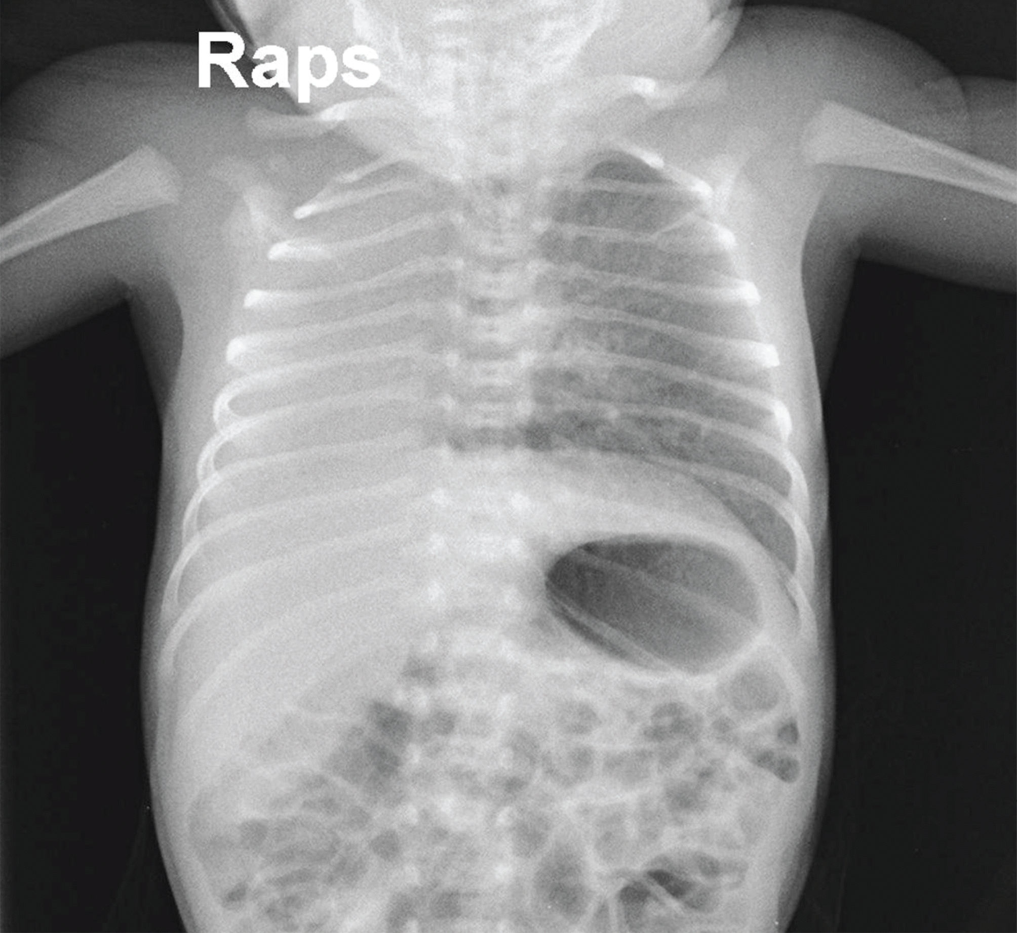
Fig. 1.
Transverse axial ultrasonography image at 35 gestational weeks shows dextroposition of the heart in the absence of intrathoracic mass. The septal axis is normal. The heart is shifted into the right chest owing to a right lung hypoplasia or agenesis.


 XML Download