

 PDF
PDF ePub
ePub Citation
 Print
Print


Abstract
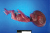
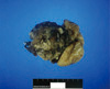
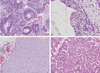
Congenital intracranial teratoma is a very rare kind of tumor. A fetus was diagnosed with a congenital intracranial teratoma and hydrocephalus at 20 weeks' gestation. On prenatal ultrasonography, the fetus showed a severe macrocephalic hydrocephalus, along with a huge, heterogenous intracranial mass in the posterior fossa. After fetal cephalocentesis was performed at 21 weeks' gestation, the pregnancy was terminated vaginally without any complication. On the postmortem examination, the pathologic report revealed an intracranial immature teratoma. We report a case with a brief review of the literature.
Figures and Tables
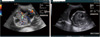
Fig. 1
Prenatal ultrasonographic findings of the cranial transverse plane. Showing about 7.1 cm sized echogenic intracranial mass with ablation of normal brain structure.
References
1. Buetow PC, Smirniotopoulos JG, Done S. Congenital brain tumors: a review of 45 cases. AJR Am J Roentgenol. 1990. 155:587–593.
2. Isaacs H. Fetal brain tumors: a review of 154 cases. Am J Perinatol. 2009. 26:453–466.
3. Inwald D, Kempley S, Hird M. Congenital primitive neuroectodermal tumour presenting as obstructed labour. Arch Dis Child Fetal Neonatal Ed. 1998. 78:F222–F224.
4. Köksal Y, Varan A, Akalan N, Bostanci A, Cila A, Söylemezoglu F, et al. Congenital cerebellar primitive neuroectodermal tumor in a newborn. Am J Perinatol. 2006. 23:173–176.
5. Kim YS, Lee KY, Kang CS, Shim SI, Kim SM. Congenital intracranial teratoma with extension into oral cavity: an autopsy case. Korean J Pathol. 1990. 24:326–330.
6. Schlembach D, Bornemann A, Rupprecht T, Beinder E. Fetal intracranial tumors detected by ultrasound: a report of two cases and review of the literature. Ultrasound Obstet Gynecol. 1999. 14:407–418.
7. Pinto V, Meo F, Loiudice L, D'Addario V. Prenatal sonographic imaging of an immature intracranial teratoma. Fetal Diagn Ther. 1999. 14:220–222.
8. Woodward PJ, Sohaey R, Kennedy A, Koeller KK. From the archives of the AFIP: a comprehensive review of fetal tumors with pathologic correlation. Radiographics. 2005. 25:215–242.
9. Schwartz S, Raffel LJ, Sun CC, Waters E. An unusual mosaic karyotype detected through prenatal diagnosis with duplication of 1q and 19p and associated teratoma development. Teratology. 1992. 46:399–404.
 XML Download