

 PDF
PDF ePub
ePub Citation
 Print
Print


Systemic lupus erythematosus (SLE) is a chronic autoimmune disease that influences pregnancies in women with normal fecundity. As the treatment of SLE has improved, more women with this disease are able to become pregnant. Pregnancy outcomes have improved dramatically over the last 40 years, with the pregnancy loss rate falling from 43% in the 1960s to 17% by 2000 [1]. Pregnancy changes affecting disease severity can be attributed to placental or maternal hormones, increased circulation, increased fluid volume, metabolic rate, hemodilution, circulating fetal cells, or other factors. While the majority of lupus pregnancies result in apparently healthy babies, lupus pregnancies are associated with a higher risk of fetal loss, premature birth, intrauterine growth retardation, neonatal lupus, and congenital heart block [2]. The rate of early pregnancy loss is approximately twice that of uncomplicated pregnancies. The rate of preterm delivery ranges from 5% to 46% [3]. Women with SLE have complicated pregnancies; one-third will result in a cesarean section, one-third will have preterm births, and >20% will be complicated by pre-eclampsia [4,5]. In addition, many medications used to treat lupus may increase the risk of birth defects [6]. The pregnancy in women with lupus must be considered to be high risk. The activity of lupus can be assessed by the C3/C4 values and anti-dsDNA titers. Although the calculation of a SLE disease activity index is standard practice, an index has not been developed for pregnant patients [7].
The medical records of 93 women diagnosed with lupus who received antepartum care at the Hanyang University Medical Center Hospital Obstetrics and Gynecology and Hanyang University Medical Center Annex Rheumatic Hospital were reviewed. The C3/C4 levels, anti-dsDNA titers, and autoimmune target test (AITT) numerical values were retrospectively analyzed to determine whether or not a correlation existed with the prognosis of lupus pregnancies.
Materials and Methods
From January 2001 to December 2008, 73 gravidas with lupus received care at Hanyang University Medical Center Obstetrics and Gynecology and Hanyang University Medical Center Rheumatic Hospital. A total of 93 pregnancies were confirmed. The diagnosis of lupus was established according to the lupus classification scheme of the American College of Rheumatology (1997). The progression of lupus, drugs taken during pregnancy, pregnancy outcomes (including abortions and stillbirths), maternal complications (pre-eclampsia, oligohydramnios, intrauterine growth retardation, early amniorrhexis, and premature labor, Raynaud syndrome, lupus nephritis, myocarditis, lung disease, hematologic disease), neonatal complications (infantile cardiac disfuntion, cerebral palsy, congenital hydronephrosis, vertebral body anomalies, anal atresia, tracheoesophageal fistula, and renal dysplasia [VATER] syndrome), hemoglobin, leucocytes, erythrocyte sedimentation rate (ESR), C-reactive protein (CRP), anti-dsDNA antibody titer (Farr assay), C3/C4 levels (radial immunodiffusion), and AITT values were determined.
Neonates were evaluated for anti-Sjögren's syndrome A (anti-SSA) (Ro) and anti-Sjögren's syndrome B (anti-SSB) (La) antibody titers, neonatal intensive care unit (NICU) admission, and the presence of disease. SPSS ver. 14.0 (SPSS Inc., Chicago, IL, USA) was used for statistical analysis. The average and standard deviation are shown. Chi-square test and Pearson correlation test were used for statistical analysis. The significance level was set at 0.05.
Results
1. Characteristics of the patients
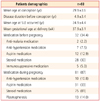
Ninety-three pregnancies in 73 patients were analyzed. The average age at the time of pregnancy was 29.9±3.7 years and the pre-gestation lupus duration of disease was 4.9±3.4 years. The average age of lupus onset was 24.9±4.4 years. The average duration of pregnancy was 37.9±3.2 weeks. Medications for the treatment of lupus were used prior to pregnancy in 34.4% of the pregnancies (32 of 93), as follows: anti-malarial agents, 2 cases (2.2%); anti-hypertensive drugs, 7 cases (7.5%); aspirin, 12 cases (12.9%); steroids, 28 cases (30.1%); and immunosuppressants, 5 cases (5.4%). Medications for lupus during pregnancy were taken 87% of pregnancies (81 of 93), as follows: anti-hypertensive drugs, 10 cases (10.8%); aspirin, 31 cases (33.3%); and steroids, 75 cases (80.6%). Plasma exchange was performed in 13 cases (14.0%) (Table 1).
2. Pregnancy outcome
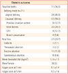
Among the 93 pregnancies, there were 22 pregnancy losses (23.7%); there were 8 spontaneous abortions (8.6%) and 1 stillbirth (1.1%). Eleven abortions (11.8%) were self-induced due to a fear of drug-induced deformities. Two pregnancies (2.2%) were terminated for medical reasons due to an exacerbation of lupus. Of the 71 pregnancies (76.3%) which continued to birth, there were 29 vaginal deliveries (31.2%) and 42 caesarean sections (45.2%). The indications for cesarean section were repeat cesarean section in 9 cases (9.7%), fetal distress in 20 cases (21.5%), cephalopelvic disproportion in 9 cases (9.7%), and breech presentation in 4 cases (4.3%). The 1 and 5 minute Apgar scores were 6.1±1.4 and 8.1±1.5, respectively (Table 2).
3. Pregnancy complications
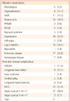
Of 71 pregnancies >20 weeks gestation, the following complications occurred: pre-eclampsia, 3 cases (4.2%); oligohydramnios, 8 cases (11.3%); preterm birth, 14 cases (23.9%); preterm premature rupture of membranes, 4 cases (5.6%); and amniorrhexis, 5 cases (7.0%). Maternal complications during pregnancy were as follows: Raynaud syndrome, 5 cases (5.4%); lupus nephritis, 10 cases (10.8%); myocarditis, 1 case (1.0%); lung disease, 17 cases (18.3%); and hematologic disease, 7 cases (7.5%).
Fetal and Neonatal complications were as follows: stillbirth, 1 case (1.4%); congenital heart defect, 9 cases (12.7%); cerebral palsy, 2 cases (2.8%); congenital hydronephrosis, 4 cases (5.6%); and VATER syndrome, 1 case (1.4%) (Table 3).
4. Activity of lupus disease during pregnancy
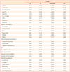
The C3 level was correlated with leukopenia, serum CRP increment, hematuria, puerperal hypertension, and preterm premature rupture of membranes. The C4 level was correlated with proteinuria, hematuria, puerperal hematologic disease, and neonatal intensive care unit admission. The anti-dsDNA titer was correlated with the serum CRP increment, oligohydramnios, and neonatal anti-SSB (La) antibody titer. The AITT was correlated with a high ESR value and 1 and 5 minute Apgar scores (Table 4).
Discussion
For gravidas with lupus, the risk of abortion, hypertension, and embryo deformity by a therapeutic agent is higher compared to healthy gravidas. Lupus flares can occur at any time during pregnancy, as well as several months after delivery [8]. Increasing doses of estrogen, as occur in pregnancy, promote physiologic and immunologic changes associated with increased lupus activity [9]. More recent studies have shown a 2-3-fold increase in SLE activity during pregnancy [10]. Fortunately, the majority of gravidas do not have severe SLE activity. In most studies, skin, joints, and constitutional symptoms are most commonly reported.
The timing of lupus activity affects the pregnancy loss rate, with activity early in pregnancy being the most clinically significant. Proteinuria, thrombocytopenia, and hypertension in the first trimester are independent risk factors for pregnancy loss [8]. Overall, approximately 20% of pregnancies in women with SLE will end with a miscarriage or stillbirth [4]. In the current study, the abortion and stillbirth rate was 23.7%. There were 11 cases (11.8%) in which curettage was self-performed due to fear of embryo deformity by a drug. The two most important risk factors for pregnancy loss are increased lupus activity and anti-phospholipid (aPL) syndrome [8]. The pathogenic role of aPL was clearly shown in experimental animals when infused during pregnancy, develop placental insufficiency, and miscarriages [11]. In addition, in vitro aPL were shown to bind trophoblastic cells and to impair the function of trophoblastic cells [12]. We also described a fetal stroke associated with maternal aPL that was detected by ultrasound and computerized tomography scan at 2 months of age in the cerebral artery territory, likely due to an intrauterine event [13]. Neonates with cerebral palsy occurred in 2 cases (2.8%).
Risk factors for preterm birth include lupus activity before and during pregnancy, higher prednisone dose, and hypertension [8]. The inflammation associated with chorioamnitis is postulated to promote dissolution of the amniotic sac, ripening of the cervix, and uterine contractions, which all lead to preterm birth [8]. In the current study, premature labor occurred in 14 cases (23.9%) of gravidas with lupus.
On average, 9.4% of all SLE pregnancy cohort births were small for gestational age (SGA), which is comparable to what would be expected in the general population [4]. Placental studies report a higher incidence of thrombosis among pregnancies affected by SLE [14]. In the current study, 11 cases (15.5%) were SGA.
Women at risk for pre-eclampsia include the following: first pregnancy; a history of pre-eclampsia or renal disease; active SLE at the time of conception; positive anti-dsDNA or anti-ribonucleoprotein antibodies; low complement activity; obesity; and/or hypertension [15]. Among lupus pregnancy cohorts, the rate of pre-eclampsia ranges from 13% to 35% [16]. Several experimental markers for pre-eclampsia, including soluble FMS-like tyrosine kinase (sFlt-1) and placental growth factor, have been found to correspond to preeclampsia in lupus patients as in women with SLE [17]. In The current study, pre-eclampsia occurred in 3 cases (4.2%).
The comparison of this research result and preexistance reports summarized in Table 5. When compared with previous studies in Korea, the frequency of stillbirth and preterm delivery is decreased. And the live birth rate is maintained.
There is no evidence that prophylactic steroids lower the frequency of flares, but there are significant adverse effects during pregnancy, including premature rupture of membranes, infections, intrauterine growth restriction, hypertension, gestational diabetes, osteoporosis, and avascular necrosis [18]. Prednisone and prednisolone are inactivated by placenta hydroxylases and <10% of the mother blood level can reach the fetus. In contrast, dexamethasone and betamethasone cannot be inactivated and treat the fetus (e.g., when incomplete congenital heart block is diagnosed) [19]. Some data suggest that prolonged fetal exposure to dexamethasone may impair cerebral development [20]. Anti-malarial drugs are widely used in autoimmune rheumatic syndromes because of their beneficial effects on skin and joints. In addition, they can lower cholesterol and lipids levels and can exert anti-aggregant activity. Drugs that are considered to be safe in pregnancy include prednisolone, azathioprine, cyclosporin A, and hydroxychloroquine. Hydroxychloroquine has been used increasingly in pregnancy with success and the published data are promising [21]. Methotrexate, mycophenolate mofetil, and cyclophosphamide are teratogenic and should be stopped at least 3 months prior to conception [22].
Nearly 20% of SLE patients are diagnosed during childhood. Although the clinical presentation and immunologic findings of the disease are similar to adults, children usually develop a more severe illness at onset with higher rates of organ involvement [23]. As with most autoimmune diseases in adults, SLE occurs more commonly in females and the role of sex hormones in disease development is recognised [23]. An erythematous skin rash with a predilection for the scalp and periorbital region, most often apparent in the first 8 weeks after birth, is also linked strongly to these maternal antibodies and to antibodies against U1 ribonucleic protein [24]. Maternal anti-Ro and anti-La antibodies may cause congenital heart block in 2% of babies [25]. Congenital heart block occurs between 18 and 30 weeks gestation, and fetal echocardiography should be performed during this period to facilitate early detection. Once detected, complete heart block cannot be reversed, but there are reports of second-degree heart block reverting to first-degree heart block after dexamethasone therapy [26]. Some studies have suggested that exposure to anti-Ro is associated with a higher prevalence of developmental dyslexia [27].
The comprehensive care of lupus patients should include a discussion of the safety and efficacy of available contraceptive options. Barrier contraceptives are safe for all women with SLE [28], but have 1-year failure rates with typical use that range from 15% to 32% [29]. Estrogen-containing contraceptives, which have 1-year failure rates of 8% with typical use [29], are safe for women who do not have aPL or who have stable lupus disease activity [30], but increase health risks for women with vascular disease. Use of the levonorgestrel-containing intrauterine device, a reversible method more effective than tubal sterilization [29], should therefore be strongly considered by women with SLE. The 1-year failure rate with typical use of a levonorgestrel-containing IUD is 0.1% [29].
The activity of the lupus can be assessed by the C3/C4 level and anti-dsDNA titer. C3 and C4 may be decreased with increased lupus activity because these proteins are consumed in the inflammatory process [31]. In pregnancy, however, the complement levels may increase 10-50% in response to increased hepatic protein synthesis [32]. During pregnancy, C3 and C4 may rise to supranormal levels, and thus a flare with complement activation may occur despite apparently normal levels of C3 and C4. Conversely, C3 and C4 may be low in the absence of a flare, probably due to synthetic defects. However, if C3 or C4 levels drop by >25%, this may be reasonably ascribed to disease activity [33]. Therefore, the utility of complement measurement in pregnancy is unclear. However, the combination of low complement levels and high-activity lupus leads to a 3-5-fold increase in pregnancy loss and preterm birth [34].
The anti-dsDNA titer is very sensitive for the diagnosis of lupus and can be indicative of increased lupus activity, especially in the kidney [8]. A rising dsDNA titer during pregnancy may correspond to increasing lupus activity; however, this antibody does not predict pregnancy outcomes. Instead, the combination of a positive anti-dsDNA titer and highly active SLE contribute toward a 4-6-fold increase in perinatal mortality and a 2-3-fold decrease in full-term birth [34].
Lupus erythematosus (LE) phenomenon was found in lupus patient's blood. LE cell test was the first autoimmune disease test of using this phenomenon that showed lower sensitivity and specificity. LE phenomenon came to be revealed in subsequent research that was generated by the reaction of various antibodies about the nuclear component. Associated with rheumatoid arthritis, a new self-antibodies (anti-microtubule organizing center and anti-Gim) was observed. So HEp-2 cell using the conventional anti-nuclear antibody (ANA) test is currently being used as a standard test. However AITT uses the macrophage cell line (IT-1) as a substrate that is wider than the ANA test in clinical applications.
The ESR is unreliable in pregnancy because it increases significantly in normal pregnancy [8]. In non-pregnant SLE patients, CRP may increase with a lupus flare. The use of CRP has not been systematically tested in SLE pregnancies [8]. A serum creatinine level >140 mmol/L is associated with a 50% pregnancy loss and this increases to 80% if the level is >400 mmol/L [28].
In this study, complement C3 levels were statistically significant in hematuria, leukopenia, hypertension, high serum CRP levels, preterm premature rupture of membranes. Complement C4 levels were statistically significant in kidney disease status, hematologic diseases, NICU. Anti-dsDNA were statistically significant in oligohydramniosd and neonatal and anti-SSB (La) antibody detection. It is helpful to predict neonatal diseases. AITT is statistically significant in a high ESR values and Apgar score. This helps to predict state of the newborn immediately after birth .
In conclusion, pregnancy rates in women with lupus disease is increasing, but systematic prenatal care improves pregnancy outcome. Still, pregnant women are concerned with fetal malformations caused by lupus drugs. Therefore, Abortion rates are higher. So an accurate perception of drug treatment and the correct method of contraception is necessary to reduce unnecessary abortion.
Complement C3/C4 tests and anti-dsDNA tests with the addition of AITT lupus can be improved prenatal care for pregnant women.
 XML Download